. 2009 Feb;91 Suppl 1(Suppl 1):95-101.
doi: 10.2106/JBJS.H.01408.

Gait mechanics influence healthy cartilage morphology and osteoarthritis of the knee

Affiliations

Gait mechanics influence healthy cartilage morphology and osteoarthritis of the knee

Thomas P Andriacchi et al. J Bone Joint Surg Am. 2009 Feb.

Abstract

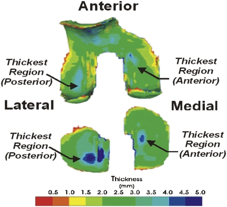
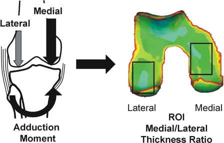
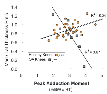
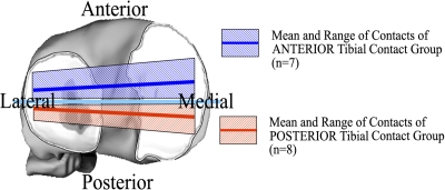
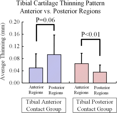
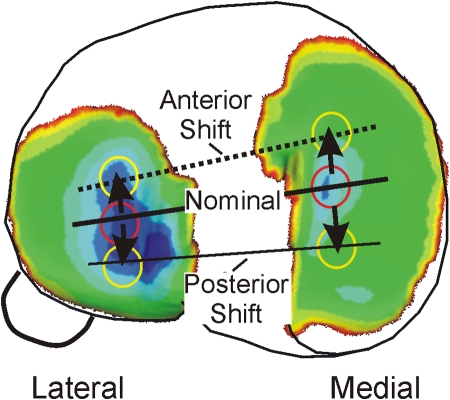
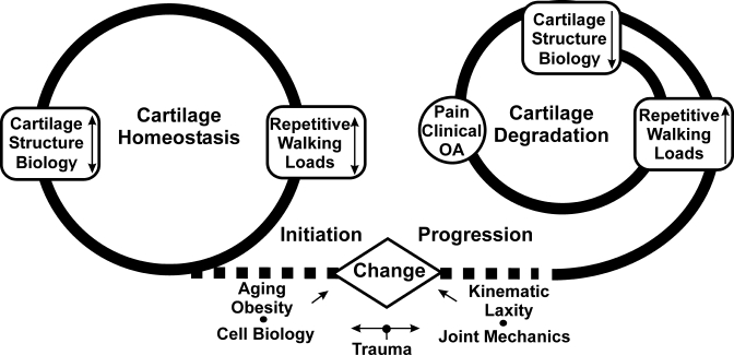
The response of healthy and diseased cartilage of the knee to the mechanics of walking is examined, with the goal of providing insight into the relationship between the kinematics and kinetics of the knee during walking and the maintenance of cartilage health. The combination of information from three-dimensional thickness models of cartilage derived from magnetic resonance imaging and the analysis of the interaction between load at the knee and kinematic changes during walking associated with loss of the anterior cruciate ligament demonstrated the importance of considering walking mechanics as an important factor in the initiation and progression of osteoarthritis. In particular, this material suggests that knee cartilage becomes conditioned to loading and to the large number of repetitive cycles of loading that occur during walking and that healthy cartilage homeostasis is maintained as long as there are no changes to the normal patterns of locomotion, the structure of the knee joint, or cartilage biology. Thus, there is the potential for a degenerative pathway to be initiated when a condition such as anterior cruciate ligament injury causes the repetitive loading during walking to shift to a new location. The sensitivity of cartilage to the kinematic changes is illustrated with the anterior cruciate ligament-deficient knee and the regional variations in cartilage morphology. The material presented here supports the conclusion that individual variations in the range of loading and kinematics at the knee during walking can have a profound influence on the initiation and progression of osteoarthritis of the knee.


Figures

Fig. 1
Fig. 1
A typical three-dimensional color thickness map of femoral and tibial cartilage. The thickest regions of the tibial and femoral weight-bearing regions (the primary weight-bearing regions during walking) are aligned when the knee is near full extension.
Fig. 2-A
Fig. 2-A
The adduction moment at the knee is associated with the medial-to-lateral load distribution at the knee. The corresponding medial and lateral femoral cartilage load-bearing regions of interest (ROI) are designated by rectangles on the color thickness map of the femoral cartilage.
Fig. 2-B
Fig. 2-B
The ratio of the medial thickness to lateral thickness in the load-bearing regions of healthy knees (represented by orange circles and dashed linear regression line, R = 0.26) during walking is greater for individuals who walk with a higher adduction moment (normalized by subject body weight × height [BW × HT]), while the relative medial thickness is less in patients who have osteoarthritis of the knee (OA) (represented by grey squares and solid linear regression line, R = 0.67) and a higher adduction moment.
Fig. 3-A Fig. 3-B
Fig. 3-A Fig. 3-B
Figs. 3-A and 3-B Patients with an anterior cruciate ligament deficiency had thinning of tibial cartilage that was associated with specific kinematic changes. The patients who had a posterior tibial shift in the anterior cruciate ligament-deficient knee relative to the intact knee had significantly greater tibial cartilage thinning in anterior regions than in posterior regions (p < 0.01), and the subjects with anterior tibial shift had more thinning in posterior regions than in anterior regions. The error bars in Figure 3-B represent one standard deviation.
Fig. 3-A Fig. 3-B
Fig. 3-A Fig. 3-B
Figs. 3-A and 3-B Patients with an anterior cruciate ligament deficiency had thinning of tibial cartilage that was associated with specific kinematic changes. The patients who had a posterior tibial shift in the anterior cruciate ligament-deficient knee relative to the intact knee had significantly greater tibial cartilage thinning in anterior regions than in posterior regions (p < 0.01), and the subjects with anterior tibial shift had more thinning in posterior regions than in anterior regions. The error bars in Figure 3-B represent one standard deviation.
Fig. 4
Fig. 4
The anterior and posterior shifts shown in Figure 3-A move general contact regions to areas of thinner cartilage in the medial compartment.
Fig. 5
Fig. 5
Healthy cartilage homeostasis is maintained by the magnitude of the repetitive cyclic loads during walking, and cartilage is thicker in regions with higher loads during walking. The initiation of osteoarthritis (OA) is associated with a change (due to injury, increased laxity, neuromuscular changes, aging, or increased obesity) in the normal balance between the mechanics of walking and the cartilage biology or structure. Once cartilage starts to degrade, it responds negatively to load and the rate of progression of osteoarthritis increases with loading.

References

    1. Andriacchi TP, Mündermann A. The role of ambulatory mechanics in the initiation and progression of knee osteoarthritis. Curr Opin Rheumatol. 2006;18:514-8. - PubMed
    1. Seedhom BB. Conditioning of cartilage during normal activities is an important factor in the development of osteoarthritis. Rheumatology (Oxford). 2006;45:146-9. - PubMed
    1. Andriacchi TP, Mündermann A, Smith RL, Alexander EJ, Dyrby CO, Koo S. A framework for the in vivo pathomechanics of osteoarthritis at the knee. Ann Biomed Eng. 2004;32:447-57. - PubMed
    1. Chaudhari AM, Briant PL, Bevill SL, Koo S, Andriacchi TP. Knee kinematics, cartilage morphology, and osteoarthritis after ACL injury. Med Sci Sports Exerc. 2008;40:215-22. - PubMed
    1. Smith RL, Donlon BS, Gupta MK, Mohtai M, Das P, Carter DR, Cooke J, Gibbons G, Hutchinson N, Schurman DJ. Effects of fluid-induced shear on articular chondrocyte morphology and metabolism in vitro. J Orthop Res. 1995;13:824-31. - PubMed