. 2009 Apr 30;113(18):4289-99.
doi: 10.1182/blood-2008-08-174797. Epub 2009 Jan 30.

Caspase-8 dependent histone acetylation by a novel proteasome inhibitor, NPI-0052: a mechanism for synergy in leukemia cells

Affiliations

Caspase-8 dependent histone acetylation by a novel proteasome inhibitor, NPI-0052: a mechanism for synergy in leukemia cells

Claudia P Miller et al. Blood. .

Abstract

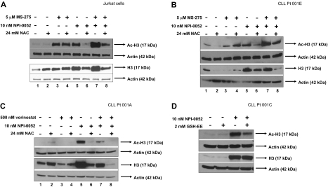
Combination studies of histone deacetylase inhibitors (HDACi) and proteasome inhibitors are providing preclinical framework to build better strategies against hematologic malignancies. Our previous work found that a novel proteasome inhibitor, NPI-0052, and HDACi synergistically induce apoptosis in leukemia cells in a caspase-8- and oxidant-dependent manner. Here we extend those observations to primary leukemia cells and identify novel mechanisms of synergy. Because the proximal targets of NPI-0052 and HDACi are inhibition of proteasome activity and histone acetylation, we initially examined those biochemical events. Increased acetylation of histone-H3 was detected in Jurkat and CLL primary cells treated with NPI-0052, alone or in combination with various HDACi (MS/SNDX-275 or vorinostat). Hyperacetylation by NPI-0052 occurred to a lesser extent in caspase-8-deficient cells and in cells treated with an antioxidant. These results indicate that NPI-0052 is eliciting caspase-8 and oxidative stress-dependent epigenetic alterations. In addition, real-time PCR revealed that MS/SNDX-275 repressed expression of the proteasomal beta5, beta2, and beta1 subunits, consequently inhibiting respective enzymatic activities. Overall, our results suggest that crosstalk by NPI-0052 and HDACi are contributing, along with caspase-8 activation and oxidative stress, to their synergistic cytotoxic effects in leukemia cells, reinforcing the potential clinical utility of combining these 2 agents.


Figures

Figure 1
Figure 1
NPI-0052 synergizes with HDAC inhibitors in primary and cultured leukemia cells. (A) Synergistic interactions between NPI-0052 and vorinostat. Jurkat cells were treated with indicated low doses of NPI-0052 and vorinostat for 24 hours. After PI cell staining, DNA fragmentation was assessed by flow cytometry. The averages of 3 independent experiments were taken to determine synergy by calculating the combination index value (CI) by isobologram analysis with Calcusyn software. A CI less than 1.0 indicates synergism. Table defines dose combinations for each data point shown and CI values. (B) HDAC inhibitors synergize more effectively with NPI-0052 than bortezomib in one AML patient sample. Mononuclear cells isolated from peripheral blood from an AML patient were treated with an HDACi/proteasome inhibitor regimen as indicated for 24 hours. DNA fragmentation was assessed after PI staining. Shown is the percent increase in the subdiploid population. CI values, indicating synergy, for each combination are shown above each bar.
Figure 2
Figure 2
Oxidative stress is elevated as a consequence of NPI-0052 and vorinostat interactions. (A) NAC partially protects from NPI-0052/vorinostat-induced apoptosis. Jurkat cells were pretreated with 24 mM NAC for 30 minutes, followed by treatment with diluent, 500 nM vorinostat, 5 nM NPI-0052, or combination of drugs for 24 hours. Apoptosis was assessed by PI staining and subsequent flow cytometric analysis. Statistical differences were calculated: *P < .05 for NPI-0052/vorinostat-treated cells compared with control (dimethyl sulfoxide [DMSO]); **P < .01 for NPI-0052/vorinostat compared with NPI-0052/vorinostat/NAC. (B) Intracellular superoxide production in Jurkat cells treated with NPI-0052/vorinostat regimen is decreased with NAC. Cells were treated as indicated for 12 hours, followed by staining with HEt and analyzed by flow cytometry. Shown are the mean fluorescence values of 3 independent experiments. (C) NPI-0052/vorinostat increases superoxide levels, which are decreased by NAC. Shown is a representative histogram of the 3 experiments performed for HEt staining (Figure 4B). Solid line represents control cells, solid bold line is 5 nM NPI-0052/500 nM vorinostat-treated cells and dashed line indicates 5 nM NPI-0052/500 nM vorinostat cells pretreated with 24 mM NAC. (D) NPI-0052 generates more superoxide levels than bortezomib in Jurkat cells. Cells were exposed to 5 nM NPI-0052, bortezomib, or diluent for 12 hours and stained with HEt. Samples were analyzed by flow cytometry. Shown are mean fluorescence values of 3 independent experiments.
Figure 3
Figure 3
HDACi, (MS-275 and vorinostat), target the proteasome. (A) MS-275 decreased mRNA expression of catalytic β5, β2, β1 proteasomal subunits. Real-time PCR analyzed expression levels of catalytic β subunits of the proteasome in Jurkat cells exposed to 5 μM MS-275 for 24 hours. Shown is the relative expression, which was calculated by first normalizing all samples to corresponding actin expression, than to control (cells treated with DMSO). *P < .05 compared with 0 hours. (B) Vorinostat decreases mRNA levels of β5, β2, β1 proteasomal subunits. Jurkat cells were treated with 500 nM vorinostat for 18 hours, and RNA was collected to analyze expression of β subunits by real-time PCR. Shown is the relative expression. *P < .01 compared with 0 hours. (C) MS-275 reduces β5, β2, β1 subunit protein expression. Jurkat cells were exposed to 5 μM MS-275, and protein lysates were collected at indicated time points. Western blot analysis and specific antibodies determined expression of β subunits and actin. Numerical values under each panel are the densitometry ratios of β subunit to actin and are normalized to 0 hours. (D) MS-275 inhibits the chymotrypsin-like and caspase-like activity of the proteasome. Jurkat cells were exposed to 5 μM MS-275 for 24 hours. The chymotrypsin-like and caspase-like activity was determined by measuring the fluorescence (amc) intensity released by the cleavage of fluorogenic substrate suc-LLVY-amc or z-LLE-amc, respectively. Proteasome activity was evaluated in RFU. Treatment with proteasome inhibitor, 10 nM NPI-0052, for 1 hour was used as a positive control. Statistical differences between 0 and 24 hours with MS-275 were determined by Student t test, *P = .002 for chymotrypsin-like and P = .004 for caspase-like. (E) Vorinostat diminishes proteasomal chymotrypsin-like and caspase-like activities. After an 18-hour exposure to 500 nM vorinostat, chymotrypsin-like and caspase-like activities in Jurkat cells were measured using fluorogenic substrates as previously described. Student t test determined statistical differences between 0 and 18 hours. (F) HDACi do not inhibit the rate-limiting activity of the proteasome in isolated proteasomes. Purified 20S proteasomes were combined with diluent, 5 μM MS-275, 500 nM vorinostat, or 10 nM NPI-0052 for 30 minutes at room temperature. The chymotrypsin-like activity was analyzed using fluorogenic substrate suc-LLVY–amc and activity was evaluated in RFU. NPI-0052 was used as a positive control.
Figure 4
Figure 4
NPI-0052 increases histone-H3 expression and its hyperacetylation. (A) NPI-0052 increases histone-H3 levels and hyperacetylation. Jurkat cells were exposed to 10 nM bortezomib, 10 nM NPI-0052, 5 μM MS-275, or a combination for 6 hours. Cell lysates were prepared and subjected to SDS polyacrylamide gel electrophoresis (SDS-PAGE) and immunobloted for acetylated (Ac)–H3, histone-H3 (H3), and actin. (B) NPI-0052 increases histone-H3 protein expression. Lysates from Jurkat cells treated with 10 nM NPI-0052 were collected at the indicated times. Western blot analyzed histone-H3 and actin expression. (C) NPI-0052 does not induce accumulation of ubiquitinated-histone-H3. Immunoprecipitation (IP) of histone H3 or p27 was performed in protein lysates from Jurkat cells that were treated with 5 μM MS-275, 10 nM NPI-0052, 200 nM NPI-0052, or a combination with MS-275 and NPI-0052 for 6 hours. After IP, samples were run on SDS-PAGE and blotted for ubiquitin. Bottom panel, protein lysates from Jurkat cells treated with the indicated compounds for 6 hours were immunoblotted for total ubiquitin protein expression.
Figure 5
Figure 5
An antioxidant attenuates hyperacetylation of histone-H3 by NPI-0052 and HDACi in cell lines and primary cells. (A) NAC decreases histone-H3 expression and hyperacetylation of histone-H3 in Jurkat cells treated with NPI-0052 and MS-275. Cells were pretreated with 24 mM NAC for 30 minutes followed with 5 μM MS-275, 10 nM NPI-0052, or combination treatment for 6 hours. Cells were harvested, lysed, and analyzed by Western blot analysis for expression of Ac-H3, histone-H3, and actin. (B) Increased histone-H3 expression and hyperacetylation as a result of NPI-0052/MS-275 interaction is decreased by NAC in CLL patient sample. Peripheral blood from patient material (patient no. 1) was separated by centrifugation with a Ficoll gradient. Isolated cells were exposed to indicated doses of NPI-0052 and MS-275 for 6 hours after exposure to 24 mM NAC. Cell lysates were analyzed for Ac-H3, histone-H3, and actin expression by Western blot analysis. (C) Hyperacetylation of histone-H3 and increased histone-H3 expression by NPI-0052/vorinostat in CLL primary cells were attenuated with NAC. Mononuclear cells were isolated from CLL patient peripheral blood (patient no. 2). Cells were treated for 6 hours with 500 nM vorinostat, 10 nM NPI-0052, or a combination after exposure to 24 mM NAC for 30 minutes. Protein lysates were prepared and Western blot analyzed expression for Ac-H3, histone-H3, and actin. (D) Supplementation with glutathione ethyl ester (GSH-EE) reduces hyperacetylation of histone-H3 by NPI-0052 in primary cells. Isolated CLL primary cells (patient no. 3) were pretreated with 2 mM GSH-EE for 1 hour, followed by exposure to 10 nM NPI-0052 or diluent for 6 hours. Protein lysates were prepared and analyzed by Western blot analysis for Ac-H3, histone-H3, and actin expression.
Figure 6
Figure 6
Caspase-8 mediates ROS production, hyperacetylation of histone-H3, and caspase-3 activation by NPI-0052/HDACi. (A) Decreased superoxide levels in a caspase-8–deficient cell line. Jurkat cells and caspase-8–deficient Jurkat cells (I9.2) were treated with 5 μM MS-275, 10 nM NPI-0052, or a combination for 12 hours. Superoxide levels were measured by staining cells with HEt and subsequent flow cytometric analysis on the FL-3 channel. Shown are mean fluorescence values for 4 independent experiments. (B) Lower superoxide levels are detected in caspase-8–deficient cells with vorinostat/NPI-0052 treatment. I9.2 and Jurkat cells were exposed to 500 nM vorinostat, 10 nM NPI-0052, or a combination for 12 hours. Dihydroethidium staining was used to detect superoxide levels. Shown are mean fluorescence values for 4 independent experiments. (C) Caspase-8 plays a role in the hyperacetylation of histone-H3 by NPI-0052. Top panel, the I9.2 (caspase-8–deficient) cells were incubated with 24 mM NAC for 30 minutes, before 5 μM MS-275, 10 nM NPI-0052, or a combination for a total of 6 hours. Cell lysates were analyzed by Western blot analysis for Ac-H3 and actin. Bottom left panel, protein lysates from Jurkat and I9.2 cells treated with 10 nM NPI-0052, 5 mM MS-275, or diluent for 6 hours were analyzed for Ac-H3 and histone H3 protein expression by Western blot analysis. Bottom right panel, protein lysates from I9.2 cells and I9.2/caspase-8/EGFP-transfected cells exposed to DMSO or 10 nM NPI-0052 for 6 hours were evaluated for Ac-H3, caspase-8, and actin protein expression by Western blot analysis. (D) Caspase-8 mediates caspase-3 activation by NPI-0052/vorinostat regimen. Caspase-3 activity was measured in Jurkat and I9.2 cells exposed to DMSO, 500 nM vorinostat, 10 nM NPI-0052, or the combination for 8 hours. *P < .001 compared control or either agent alone; †P < .001 compared with Jurkat cells incubated with NPI-0052/vorinostat.

References

    1. Mehta PA, Davies SM. Allogeneic transplantation for childhood ALL. Bone Marrow Transplant. 2008;41:133–139. - PubMed
    1. Kassim AA, Chinratanalab W, Ferrara JL, Mineishi S. Reduced-intensity allogeneic hematopoietic stem cell transplantation for acute leukemias: ‘what is the best recipe?’. Bone Marrow Transplant. 2005;36:565–574. - PubMed
    1. O'Brien SM, Kantarjian HM, Cortes J, et al. Results of the fludarabine and cyclophosphamide combination regimen in chronic lymphocytic leukemia. J Clin Oncol. 2001;19:1414–1420. - PubMed
    1. Hijiya N, Hudson MM, Lensing S, et al. Cumulative incidence of secondary neoplasms as a first event after childhood acute lymphoblastic leukemia. JAMA. 2007;297:1207–1215. - PubMed
    1. Lipshultz SE, Lipsitz SR, Sallan SE, et al. Chronic progressive cardiac dysfunction years after doxorubicin therapy for childhood acute lymphoblastic leukemia. J Clin Oncol. 2005;23:2629–2636. - PubMed

Publication types

MeSH terms