

In vivo imaging of the mouse model of X-linked juvenile retinoschisis with fourier domain optical coherence tomography
- PMID: 19182246
- PMCID: PMC2693243
- DOI: 10.1167/iovs.08-2542
In vivo imaging of the mouse model of X-linked juvenile retinoschisis with fourier domain optical coherence tomography
Abstract
Purpose: The purpose of this study was to investigate Fourier domain optical coherence tomography (FD OCT) as a noninvasive tool for retinal imaging in the Rs1h-knockout mouse (model for X-linked juvenile retinoschisis).
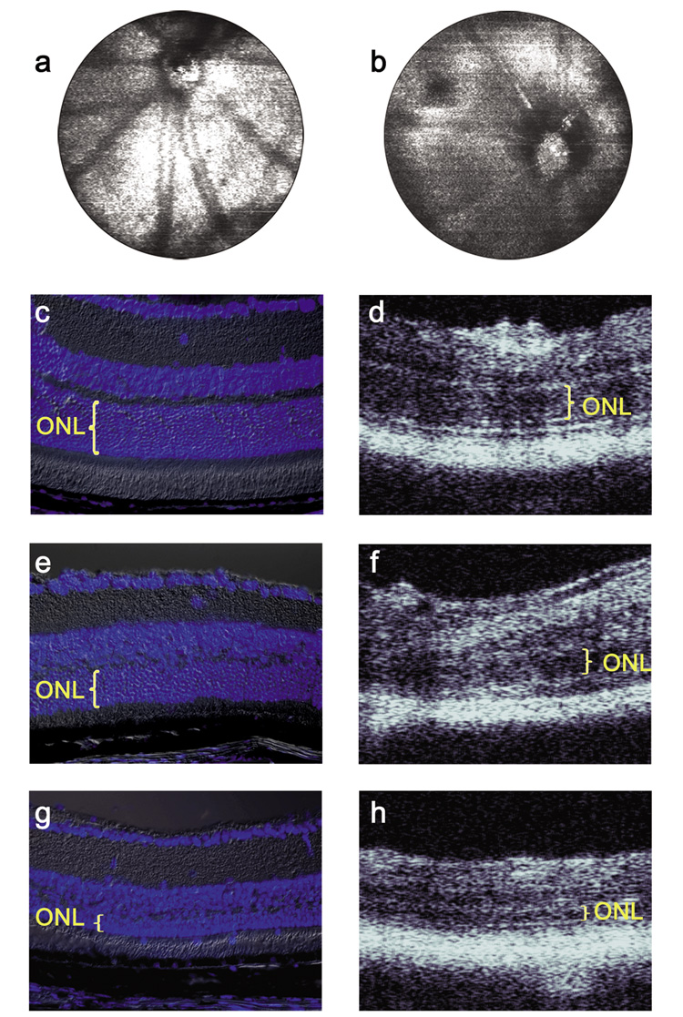
Methods: A prototype spectrometer-based FD OCT system was used in combination with a custom optical beam-scanning platform. Images of the retinas from wild-type and Rs1h-knockout mice were acquired noninvasively with FD OCT with the specimen anesthetized. At the completion of the noninvasive FD OCT imaging, invasive retinal cross-sectional images (histology) were acquired from a nearby region for comparison to the FD OCT images.
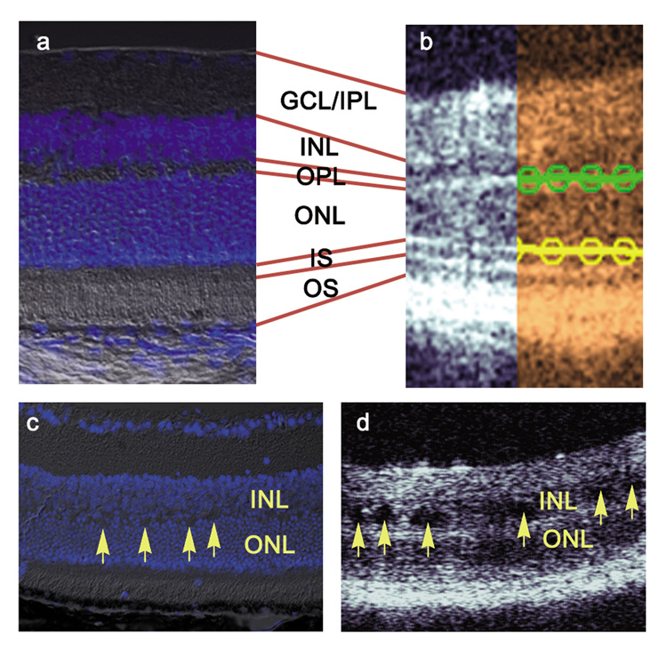
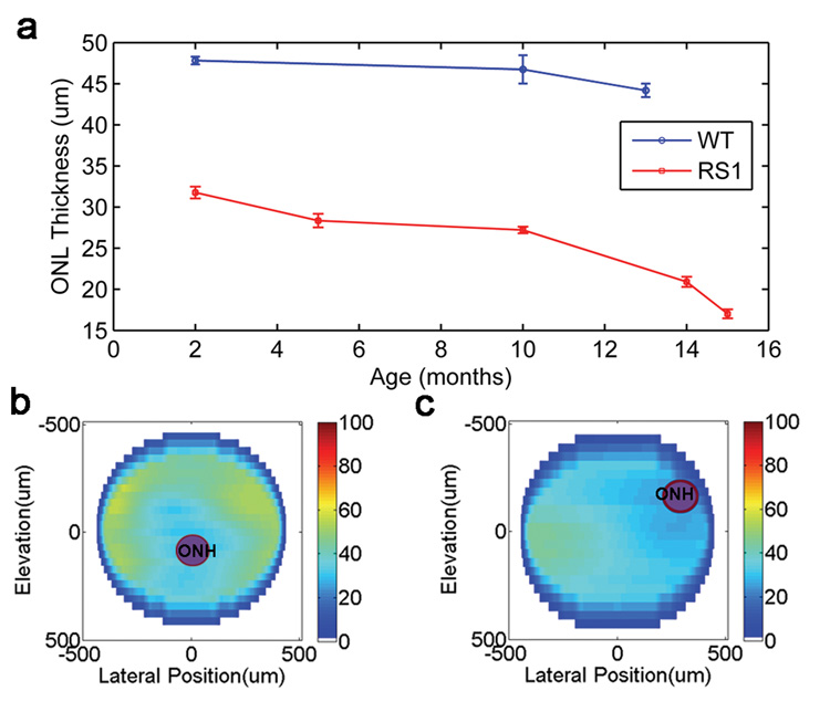
Results: The retinal layers were identifiable in the FD OCT images, permitting delineation and thickness measurement of the outer nuclear layer (ONL). During FD OCT in vivo imaging of the Rs1h-knockout mouse, holes were observed in the inner nuclear layer (INL), and retinal cell disorganization was observed as a change in the backscattering intensity profile. Comparison of the ONL measurements acquired noninvasively with FD OCT to measurements taken using histology at nearby locations showed a degeneration of roughly 30% of the ONL by the age of 2 months in Rs1h-knockout mice relative to wild-type.
Conclusions: FD OCT was demonstrated to be effective for noninvasive imaging of retinal degeneration and observation of retinal holes in Rs1h-knockout mice.
Figures

Similar articles
-
Retinoschisin gene therapy and natural history in the Rs1h-KO mouse: long-term rescue from retinal degeneration.Invest Ophthalmol Vis Sci. 2007 Aug;48(8):3837-45. doi: 10.1167/iovs.07-0203. Invest Ophthalmol Vis Sci. 2007. PMID: 17652759
-
RS-1 Gene Delivery to an Adult Rs1h Knockout Mouse Model Restores ERG b-Wave with Reversal of the Electronegative Waveform of X-Linked Retinoschisis.Invest Ophthalmol Vis Sci. 2004 Sep;45(9):3279-85. doi: 10.1167/iovs.04-0576. Invest Ophthalmol Vis Sci. 2004. PMID: 15326152
-
Fourier domain optical coherence tomography as a noninvasive means for in vivo detection of retinal degeneration in Xenopus laevis tadpoles.Invest Ophthalmol Vis Sci. 2010 Feb;51(2):1066-70. doi: 10.1167/iovs.09-4260. Epub 2009 Sep 9. Invest Ophthalmol Vis Sci. 2010. PMID: 19741241
-
Of men and mice: Human X-linked retinoschisis and fidelity in mouse modeling.Prog Retin Eye Res. 2022 Mar;87:100999. doi: 10.1016/j.preteyeres.2021.100999. Epub 2021 Aug 11. Prog Retin Eye Res. 2022. PMID: 34390869 Review.
-
Biology of retinoschisin.Adv Exp Med Biol. 2012;723:513-8. doi: 10.1007/978-1-4614-0631-0_64. Adv Exp Med Biol. 2012. PMID: 22183371 Free PMC article. Review. No abstract available.
Cited by
-
Evaluation of inner retinal thickness around the optic disc using optical coherence tomography of a rodent model of nonarteritic ischemic optic neuropathy.Jpn J Ophthalmol. 2013 May;57(3):327-32. doi: 10.1007/s10384-012-0195-7. Epub 2012 Oct 10. Jpn J Ophthalmol. 2013. PMID: 23053634
-
X-linked juvenile retinoschisis: clinical diagnosis, genetic analysis, and molecular mechanisms.Prog Retin Eye Res. 2012 May;31(3):195-212. doi: 10.1016/j.preteyeres.2011.12.002. Epub 2012 Jan 3. Prog Retin Eye Res. 2012. PMID: 22245536 Free PMC article. Review.
-
Morphometric and Microstructural Changes During Murine Retinal Development Characterized Using In Vivo Optical Coherence Tomography.Invest Ophthalmol Vis Sci. 2021 Oct 4;62(13):20. doi: 10.1167/iovs.62.13.20. Invest Ophthalmol Vis Sci. 2021. PMID: 34698774 Free PMC article.
-
X-Linked Retinoschisis: Phenotypic Variability in a Chinese Family.Sci Rep. 2016 Jan 29;6:20118. doi: 10.1038/srep20118. Sci Rep. 2016. PMID: 26823236 Free PMC article. Clinical Trial.
-
Adaptive optics optical coherence tomography for in vivo mouse retinal imaging.J Biomed Opt. 2013 May;18(5):56007. doi: 10.1117/1.JBO.18.5.056007. J Biomed Opt. 2013. PMID: 23644903 Free PMC article.
References
-
- Fercher AF, Hitzenberger CK, Kamp G, Elzaiat SY. Measurement of Intraocular Distances by Backscattering Spectral Interferometry. Optics Communications. 1995;117(1–2):43–48.
-
- Cense B, Nassif N, Chen TC, Pierce MC, Yun SH, Park BH, Bouma BE, Tearney GJ, de Boer JF. Ultrahigh-resolution high-speed retinal imaging using spectral-domain optical coherence tomography. Optics Express. 2004;12(11):2435–2447. - PubMed
-
- Wojtkowski M, Srinivasan VJ, Ko TH, Fujimoto JG, Kowalczyk A, Duker JS. Ultrahigh-resolution, high-speed, Fourier domain optical coherence tomography and methods for dispersion compensation. Optics Express. 2004;12(11):2404–2422. - PubMed
-
- Leitgeb RA, Drexler W, Unterhuber A, Hermann B, Bajraszewski T, Le T, Stingl A, Fercher AF. Ultrahigh resolution Fourier domain optical coherence tomography. Optics Express. 2004;12(10):2156–2165. - PubMed
Publication types
MeSH terms
Substances
Grants and funding
LinkOut - more resources
Full Text Sources
Molecular Biology Databases
