

The clinical usefulness of (18)F-FDG PET/CT for the evaluation of lymph node metastasis in periorbital malignancies
- PMID: 19182496
- PMCID: PMC2647179
- DOI: 10.3348/kjr.2009.10.1.1
The clinical usefulness of (18)F-FDG PET/CT for the evaluation of lymph node metastasis in periorbital malignancies
Abstract
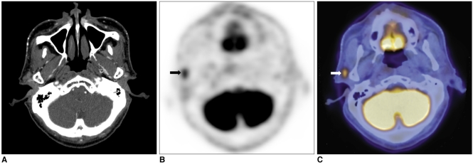
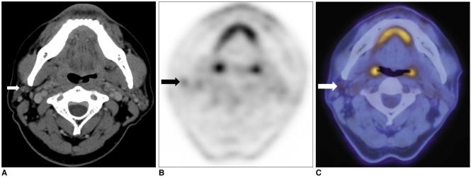
Objective: The aim of this study was to assess the clinical role of (18)F-FDG PET/CT for the evaluation of lymph node metastasis in periorbital malignancies, compared with CT alone.
Materials and methods: We analyzed eighteen PET/CT and CT scans in 15 patients with biopsy-proven periorbital malignancies. We compared the diagnostic capabilities of PET/CT and CT with regard to nodal metastasis by level-by-level analysis and by N staging prediction. The reference standards were surgical pathology (n = 7) from dissected lymph node specimens and the results from radiological follow-up (n = 11, mean 20.5 months; range 10-52 months). Moreover, any changes in patient care as prompted by PET/CT were recorded and compared with treatment planning for CT alone.
Results: PET/CT had a sensitivity of 100%, while CT had a sensitivity of 57% (p = 0.03) for nodal metastasis by level-by-level analysis. PET/CT had a specificity of 97%, positive predictive value of 93%, negative predictive value of 100%, and diagnostic accuracy of 98%, while the CT values for these same parameters were 97%, 89%, 82%, and 84%, respectively. PET/CT correctly predicted N staging with an accuracy of 100%, while CT was only 83% accurate (p = 0.01). Regarding the impact on patient care, the extent of surgery for regional lymph nodes and the treatment decision were modified by PET/CT in 39% of patients.
Conclusion: PET/CT could provide useful information in the management of regional lymph node metastases in patients with periorbital malignancies.
Figures
References
-
- Limawararut V, Leibovitch I, Sullivan T, Selva D. Periocular squamous cell carcinoma. Clin Experiment Ophthalmol. 2007;35:174–185. - PubMed
-
- Cook BE, Jr, Bartley GB. Treatment options and future prospects for the management of eyelid malignancies: an evidence-based update. Ophthalmology. 2001;108:2088–2098. - PubMed
-
- Reifler DM, Hornblass A. Squamous cell carcinoma of the eyelid. Surv Ophthalmol. 1986;30:349–365. - PubMed
-
- Kass LG, Hornblass A. Sebaceous carcinoma of the ocular adnexa. Surv Ophthalmol. 1989;33:477–490. - PubMed
