. 2009 Mar;5(3):157-65.
doi: 10.1038/nchembio.144. Epub 2009 Feb 1.

Golgicide A reveals essential roles for GBF1 in Golgi assembly and function

Affiliations

Golgicide A reveals essential roles for GBF1 in Golgi assembly and function

José B Sáenz et al. Nat Chem Biol. 2009 Mar.

Abstract

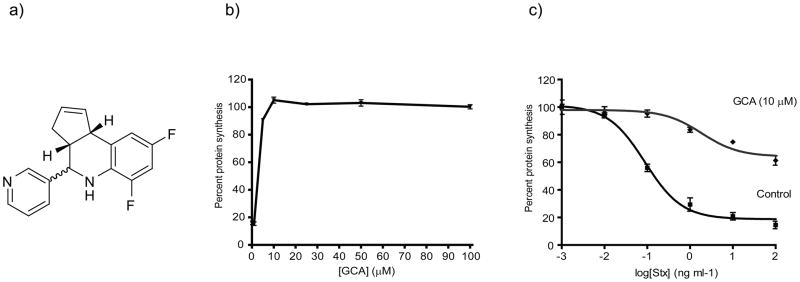
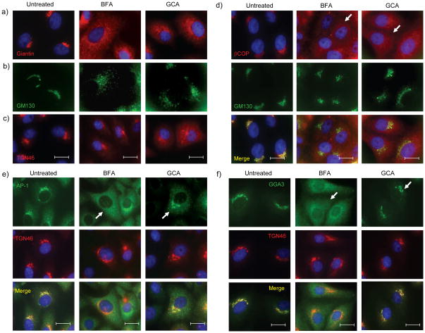
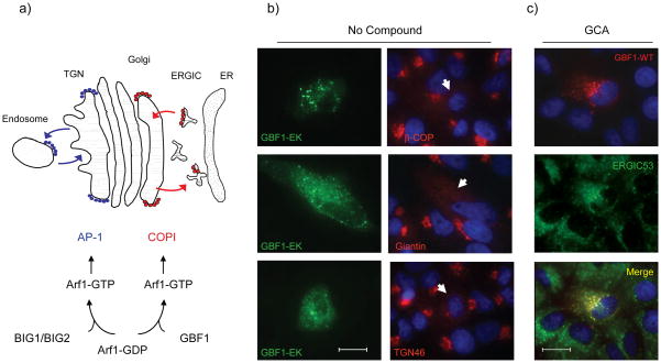
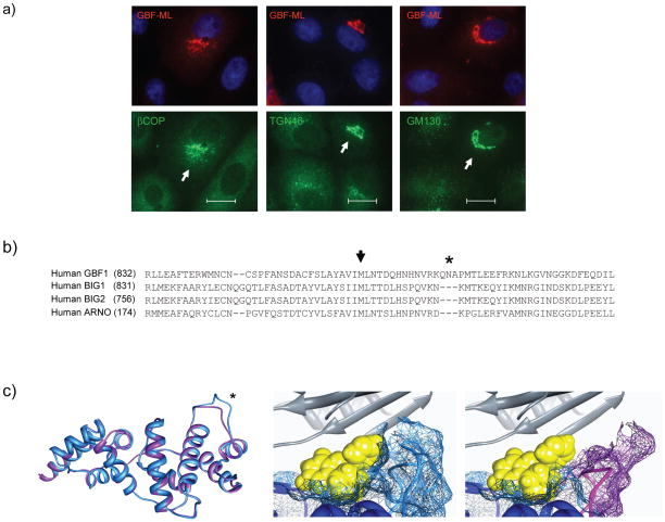
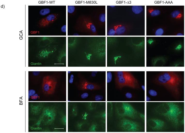
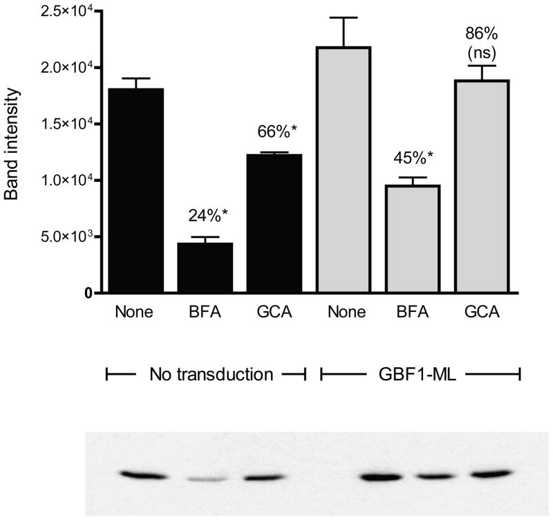
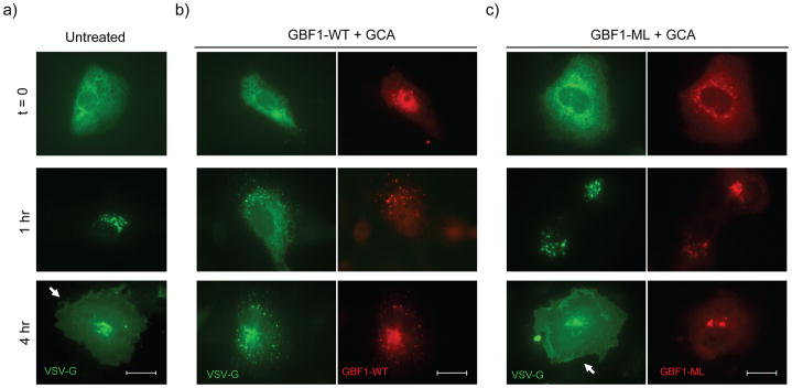
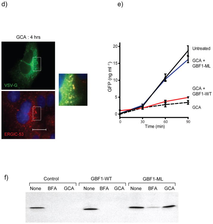
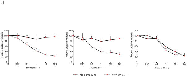
ADP ribosylation factor 1 (Arf1) plays a critical role in regulating secretory traffic and membrane transport within the Golgi of eukaryotic cells. Arf1 is activated by guanine nucleotide exchange factors (ArfGEFs), which confer spatial and temporal specificity to vesicular transport. We describe here the discovery and characterization of golgicide A, a potent, highly specific, reversible inhibitor of the cis-Golgi ArfGEF GBF1. Inhibition of GBF1 function resulted in rapid dissociation of COPI vesicle coat from Golgi membranes and subsequent disassembly of the Golgi and trans-Golgi network. Secretion of soluble and membrane-associated proteins was arrested at the endoplasmic reticulum-Golgi intermediate compartment, whereas endocytosis and recycling of transferrin were unaffected by GBF1 inhibition. Internalized shiga toxin was arrested within the endocytic compartment and was unable to reach the dispersed trans-Golgi network. Collectively, these results highlight the central role for GBF1 in coordinating bidirectional transport and maintaining structural integrity of the Golgi.


Figures

Figure 1
Figure 1
Discovery of GCA as a potent and effective inhibitor of Stx susceptibility. (a) Structure of GCA. (b) Vero cells were pretreated for 30 min at 37°C with varying concentrations of GCA, followed by incubation with Stx (1 ng/mL) for 4 h at 37°C. % Protein synthesis indicates the amount of radioactive amino acid incorporation in GCA-treated cells as a percentage of radioactive amino acid incorporation in cells lacking Stx treatment (control). Protein synthesis levels and compound-response curves were determined as described in Methods. (c) Protein synthesis levels for control (squares; no compound) or GCA-treated (triangles; 10 μM) Vero cells were determined using the radioactive amino acid incorporation assay as described in (b). % Protein synthesis is expressed as the amount of radioactive amino acid incorporation in untreated or GCA-treated cells at a given toxin concentration as a percentage of radioactive amino acid incorporation in cells lacking Stx treatment. Toxin IC50 values for GCA-treated cells were significantly increased over control cells (p<0.01; see Methods). For (a) and (b), data points (mean ±SD) represent triplicate data at the indicated compound or toxin concentrations, respectively, from one representative experiment. Stx, Shiga toxin; GCA, golgicide A.
Figure 2
Figure 2
GCA disperses medial- and cis-Golgi, inhibits COPI recruitment, and maintains localization of AP-1 and GGA3 to TGN. (a,b) Vero cells were either left untreated or treated with BFA (10 μg/mL) or GCA (10 μM) for 1 hr, and the localization of (a) giantin (medial-Golgi), (b) GM130 (cis-Golgi), and (c) TGN46 (TGN) were determined (see Methods). The distribution of medial- and cis-Golgi was similar in BFA- and GCA-treated cells, while GCA did not induce tubulation of the TGN as observed in BFA-treated cells. (d) Vero cells treated for 5 min with BFA (10 μg/mL) or GCA (10 μM) show dispersed COPI staining (arrows) that does not colocalize with the Golgi (green) as in untreated cells. (e,f) Unlike BFA, GCA maintains (e) AP-1 and (f) GGA3 association with the TGN after 5 min of treatment (see arrows). BFA, brefeldin A; blue, nuclei. White scale bars = 20 μm.
Figure 3
Figure 3
The effects of GCA are similar to expression of inactive GBF1-E794K. (a) Schematic diagram of the roles of GBF1 and BIG1/BIG2 in Arf1 activation. Arf1 in the inactive (GDP-bound) form is localized to the cytoplasm. Exchange of GDP for GTP by either BIG1/2 or GBF1 results in local Arf1 activation, membrane localization, and subsequent recruitment of vesicle coat proteins such as AP-1 or COPI. (b) Vero cells expressing the GBF1-EK mutant show dispersal of β-COP, the medial-Golgi (giantin), and the trans-Golgi network (TGN46). Arrows indicate the effect of GBF1-E794K expression on giantin, βCOP, or TGN46 localization. Blue, nuclei. (c) GBF1-WT is predominantly membrane-associated in GCA-treated cells and distributed to punctate structures that co-label with anti-ERGIC53. White scale bars = 20 μm.
Figure 4
Figure 4
GCA is selective for GBF1 (a) Expression of GBF1-M832L (GBF1-ML) results in protection from the effects of GCA. Vero cells were transiently transfected with GBF1-ML and exposed to 100 μM GCA. β-COP, TGN46, and GM130 localization were resistant to the effects of GCA. Arrows indicate cells expressing GBF1-ML. Blue, nuclei. White scale bars = 20 μm. (b) Comparison of the amino acid sequence of Sec7 domains reveals considerable divergence between GBF1 and BIG1/BIG2. Particularly notable was the presence of three extra residues in the GBF1 sequence (asterisk) (c) The GBF1 Sec7 domain (residues 693 to 887) was thread on the previously reported ARNO-Arf1-BFA complex. Only the Sec7 domains of GBF1 (blue) and ARNO (purple) are shown (left panel). The modeled tertiary structures are nearly identical, with the exception of an additional loop in the GBF1 Sec7 domain (asterisk), corresponding to the tripeptide insertion in GBF1 sequence. GCA was docked into the BFA binding pocket, revealing the compound to extend past the BFA-binding region to make contact with the GBF1 tripeptide loop (middle panel; light blue). In these images, predicted molecular surfaces are indicated by colored mesh around the ribbon backbone. The corresponding region of ARNO, BIG1 and BIG2 lacks the tripeptide (right panel, purple) and does not contact GCA. In these images, GCA is yellow and Arf1 ribbon diagram is gray. (d) Mutagenesis of the tripeptide, either by deletion of the three residues, or alteration to three alanine residues results in resistance to GCA but not BFA. Cells were transiently transfected either with GBF1-WT, GBF1-ML, GBF1-Δ3 (QNV deleted), or GBF1-AAA (QNV to AAA) then exposed to GCA (10 μM; top panel) or BFA (10 μg/ml; bottom panel) for 1 hr, and labeled with antibodies against HA and giantin. White scale bars = 20 μm.
Figure 4
Figure 4
GCA is selective for GBF1 (a) Expression of GBF1-M832L (GBF1-ML) results in protection from the effects of GCA. Vero cells were transiently transfected with GBF1-ML and exposed to 100 μM GCA. β-COP, TGN46, and GM130 localization were resistant to the effects of GCA. Arrows indicate cells expressing GBF1-ML. Blue, nuclei. White scale bars = 20 μm. (b) Comparison of the amino acid sequence of Sec7 domains reveals considerable divergence between GBF1 and BIG1/BIG2. Particularly notable was the presence of three extra residues in the GBF1 sequence (asterisk) (c) The GBF1 Sec7 domain (residues 693 to 887) was thread on the previously reported ARNO-Arf1-BFA complex. Only the Sec7 domains of GBF1 (blue) and ARNO (purple) are shown (left panel). The modeled tertiary structures are nearly identical, with the exception of an additional loop in the GBF1 Sec7 domain (asterisk), corresponding to the tripeptide insertion in GBF1 sequence. GCA was docked into the BFA binding pocket, revealing the compound to extend past the BFA-binding region to make contact with the GBF1 tripeptide loop (middle panel; light blue). In these images, predicted molecular surfaces are indicated by colored mesh around the ribbon backbone. The corresponding region of ARNO, BIG1 and BIG2 lacks the tripeptide (right panel, purple) and does not contact GCA. In these images, GCA is yellow and Arf1 ribbon diagram is gray. (d) Mutagenesis of the tripeptide, either by deletion of the three residues, or alteration to three alanine residues results in resistance to GCA but not BFA. Cells were transiently transfected either with GBF1-WT, GBF1-ML, GBF1-Δ3 (QNV deleted), or GBF1-AAA (QNV to AAA) then exposed to GCA (10 μM; top panel) or BFA (10 μg/ml; bottom panel) for 1 hr, and labeled with antibodies against HA and giantin. White scale bars = 20 μm.
Figure 5
Figure 5
GCA causes a decrease in GBF1-dependent Arf1 activation. Vero cells transduced with Arf1-V5 alone or Arf1-V5 plus GBF1-M832L-HA were exposed either to no compound (Untreated), BFA (10 μg/ml) or GCA (10 μM) for 1 hr. The cells were then lysed and extracts incubated with immobilized GST-GGA3. Bound proteins were released and separated by SDS-PAGE. Arf1-V5 was detected by Western blot and band intensity was determined using ImageJ software. Statistical analysis was performed on duplicate experiments. (*, statistically significant from untreated sample [P=0.05]; ns, not statistically significantly different from untreated sample).
Figure 6
Figure 6
GBF1 inhibition arrests secretion of membrane-anchored and soluble proteins. Trafficking of tsVSVG-GFP (green) was arrested in GCA-treated cells. Vero cells were co-transfected with tsVSVG-GFP and either (a) no additional plasmid, (b) wild-type GBF1 (GBF1-WT), or (c) the GBF1-M832L mutant (GBF1-ML). The cells were incubated at 40°C to trap tsVSVG-GFP in the ER, followed by treatment with either (a) no compound or (b,c) GCA (10 μM) prior to shifting the cells to 32°C. At various times (t = 0, 1, and 4 h) following the temperature shift, cells were fixed and labeled with anti-HA antibody (GBF1 proteins; red). Expression of the GBF1-ML mutant restored tsVSVG-GFP transit to the plasma membrane (arrow). (d) tsVSVG-GFP co-localizes with ERGIC53 in GCA-treated cells. Vero cells transfected with tsVSVG-GFP were incubated at 40°C before a shift to 32°C for 4 h in the presence of GCA (10 μM). The cells were fixed and labeled with an anti-ERGIC53 antibody (red). Inset shows magnification of boxed area and co-localization of VSVG with the ERGIC (yellow). Blue, nuclei. White scale bars = 20 μm. (e) GCA inhibits secretion of soluble cargo. Cells were transduced either with adenovirus expressing NPY-GFP alone or co-transduced with NPY-GFP plus adenovirus expressing GBF1-WT or GBF1-ML. The cells were either left untreated or were exposed to GCA (10 μM) for 1 h. At various times, the media was removed and GFP concentration assessed by ELISA (see Supplementary Methods). GCA treatment decreases NPY-GFP secretion in untransduced cells (dotted), while overexpression of GBF1-ML (blue), but not GBF1-WT (red), restores NPY-GFP secretion to levels in untransduced, untreated cells (black). (f) Sulfation of a StxB construct with a tandem of sulfation sites (StxB-SS) was used to assess trafficking from endosomes to the TGN. Control Vero cells, or those transduced with GBF1-WT or GBF1-ML were preincubated with no compound, BFA (10 μg/ml) or GCA (10 μM), then StxB-SS was added for 3 hrs in the presence of [35S]O4 as described in Supplementary Methods. StxB-SS was recovered from cell lysates and subjected to SDS-PAGE and autoradiography to detect radiolabeled toxin. (g) Inhibition of shiga toxicity by GCA is completely reversed in cells expressing GBF1-ML. Cells were either left untransduced or transduced with adenovirus expressing GBF1-WT or GBF1-ML and were exposed either to GCA (solid line) or no compound (dotted line) prior to treatment with increasing concentrations of Stx. After 4 h incubation, protein synthesis was assessed, as described in Supplementary Methods. GBF1-ML expression restores Stx susceptibility in GCA-treated cells.
Figure 6
Figure 6
GBF1 inhibition arrests secretion of membrane-anchored and soluble proteins. Trafficking of tsVSVG-GFP (green) was arrested in GCA-treated cells. Vero cells were co-transfected with tsVSVG-GFP and either (a) no additional plasmid, (b) wild-type GBF1 (GBF1-WT), or (c) the GBF1-M832L mutant (GBF1-ML). The cells were incubated at 40°C to trap tsVSVG-GFP in the ER, followed by treatment with either (a) no compound or (b,c) GCA (10 μM) prior to shifting the cells to 32°C. At various times (t = 0, 1, and 4 h) following the temperature shift, cells were fixed and labeled with anti-HA antibody (GBF1 proteins; red). Expression of the GBF1-ML mutant restored tsVSVG-GFP transit to the plasma membrane (arrow). (d) tsVSVG-GFP co-localizes with ERGIC53 in GCA-treated cells. Vero cells transfected with tsVSVG-GFP were incubated at 40°C before a shift to 32°C for 4 h in the presence of GCA (10 μM). The cells were fixed and labeled with an anti-ERGIC53 antibody (red). Inset shows magnification of boxed area and co-localization of VSVG with the ERGIC (yellow). Blue, nuclei. White scale bars = 20 μm. (e) GCA inhibits secretion of soluble cargo. Cells were transduced either with adenovirus expressing NPY-GFP alone or co-transduced with NPY-GFP plus adenovirus expressing GBF1-WT or GBF1-ML. The cells were either left untreated or were exposed to GCA (10 μM) for 1 h. At various times, the media was removed and GFP concentration assessed by ELISA (see Supplementary Methods). GCA treatment decreases NPY-GFP secretion in untransduced cells (dotted), while overexpression of GBF1-ML (blue), but not GBF1-WT (red), restores NPY-GFP secretion to levels in untransduced, untreated cells (black). (f) Sulfation of a StxB construct with a tandem of sulfation sites (StxB-SS) was used to assess trafficking from endosomes to the TGN. Control Vero cells, or those transduced with GBF1-WT or GBF1-ML were preincubated with no compound, BFA (10 μg/ml) or GCA (10 μM), then StxB-SS was added for 3 hrs in the presence of [35S]O4 as described in Supplementary Methods. StxB-SS was recovered from cell lysates and subjected to SDS-PAGE and autoradiography to detect radiolabeled toxin. (g) Inhibition of shiga toxicity by GCA is completely reversed in cells expressing GBF1-ML. Cells were either left untransduced or transduced with adenovirus expressing GBF1-WT or GBF1-ML and were exposed either to GCA (solid line) or no compound (dotted line) prior to treatment with increasing concentrations of Stx. After 4 h incubation, protein synthesis was assessed, as described in Supplementary Methods. GBF1-ML expression restores Stx susceptibility in GCA-treated cells.

Similar articles

Cited by

References

    1. Donaldson JG, Honda A, Weigert R. Multiple activities for Arf1 at the Golgi complex. Biochim Biophys Acta. 2005;1744:364–73. - PubMed
    1. Cohen LA, et al. Active Arf6 recruits ARNO/cytohesin GEFs to the PM by binding their PH domains. Mol Biol Cell. 2007;18:2244–53. - PMC - PubMed
    1. Shmuel M, et al. ARNO through its coiled-coil domain regulates endocytosis at the apical surface of polarized epithelial cells. J Biol Chem. 2006;281:13300–8. - PubMed
    1. Szul T, et al. Dissecting the role of the ARF guanine nucleotide exchange factor GBF1 in Golgi biogenesis and protein trafficking. J Cell Sci. 2007;120:3929–40. - PubMed
    1. Zhao X, et al. GBF1, a cis-Golgi and VTCs-localized ARF-GEF, is implicated in ER-to-Golgi protein traffic. J Cell Sci. 2006;119:3743–53. - PubMed

Publication types