Comparative Study
. 2009 Mar;12(3):268-76.
doi: 10.1038/nn.2254. Epub 2009 Feb 1.

Ephrin-B3 reverse signaling through Grb4 and cytoskeletal regulators mediates axon pruning

Affiliations
Comparative Study

Ephrin-B3 reverse signaling through Grb4 and cytoskeletal regulators mediates axon pruning

Nan-Jie Xu et al. Nat Neurosci. 2009 Mar.

Abstract

It has been suggested that ephrin-B proteins have receptor-like roles in the control of axon pathfinding by repulsion, although it is largely unknown how the reverse signals are coupled to downstream intracellular molecules and how they induce cytoskeletal reorganization at the axon terminal. We found that ephrin-B3 (EB3) was able to function as a repulsive guidance receptor and mediate stereotyped pruning of murine hippocampal mossy fiber axons during postnatal development. Targeted intracellular point mutants showed that axon pruning requires tyrosine phosphorylation-dependent reverse signaling and coupling to the SH2/SH3 adaptor protein Grb4 (also known as Nckbeta/Nck2). Furthermore, we found that the second SH3 domain of Grb4 is required and sufficient for axon pruning/retraction by mediating interactions with Dock180 and PAK to bring about guanine nucleotide exchange and signaling downstream of Rac, respectively. Our results reveal a previously unknown pathway that controls axon pruning and elucidate the biochemical mechanism by which ephrin-B reverse signals regulate actin dynamics to bring about the retraction of growth cones.


Figures
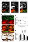
Figure 1
Figure 1
Defective hippocampal MF pruning in EB3 mutants. a, Confocal IF detects the EB3-β-gal fusion protein in calbinding-positive SPB (arrows) and IPB (arrowheads) MF axons extending from the DG towards the CA3 area in postnatal day 10 (PD10) and 10 week old adult (PW10) mice. The EphB2-β-gal fusion protein is expressed broadly above and underneath the MF bundles, but is not localized with calbindin in the MF axons. b and c, Anti-calbindin IF (b) and Timm stain (c) shows that IPB axons in 8–10 week old adult EB3−/− mice are much longer than in WT littermates (distance between arrowheads). d, Quantification of the ratio of IPB length/length from hilus to curvature of CA3 area in EB3−/− and WT littermates during postnatal development (n = 3–4 per group). e, Quantification of IPB length in EB3−/−, EB3lacZ/lacZ, and WT adult mice (n = 8–9 per group). Mean ± s.e.m. Scale bars: 300 μm in a, b and c (upper panels); 150 μm in b and c (bottom panels).
Figure 2
Figure 2
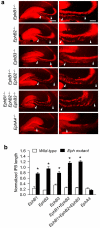
Defective hippocampal MF pruning in EphB mutants. a, Anti-calbindin IF shows long IPB axons in indicated EphB single and compound mutants at 8–10 weeks. b, Quantification of IPB length in EphB1−/− (n = 6), EphB2−/− (n = 5), EphB3−/− (n = 4), EphB1−/−; EphB2−/− (n = 6), EphB1−/−; EphB2−/−; EphB3−/− (n = 6), and EphA4−/− (n = 6) mutants (* P < 0.001). Mean ± s.e.m. Scale bars: 300 μm in a (left panels); 150 μm in a (right panels).
Figure 3
Figure 3
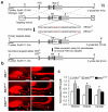
Mossy fiber pruning requires tyrosine phosphorylation of the EB3 cytoplasmic tail. a, Generation of EB3 tyrosine (Y) to phenylalanine (F) point mutations. Exons are shown as filled boxes (coding segments are dark and non-coding segments are gray), introns and 5′ and 3′ nontranscribed regions as lines, and EcoRI (E) and NsiI (N) restriction sites are indicated. The last (fifth) exon encoding EB3 amino acids 205–340 including the cytoplasmic domain was engineered to generate 3F or 5F mutations (red) following homologous recombination in ES cells and Cre-mediated excision of the loxP-flanked PGK-neo cassette after germline transmission of the initial targeted EB3neo insertions. The location of 5′ and 3′ external probes used to confirm the various recombination events by Southern blot are shown with the expected sizes indicated (see Supplementary Fig. 7). b, The IPB axons in both EB33F/3F and EB35F/5F mutants are longer than in WT mice. c, Quantification of IPB length in 8–10 week old EB33F/3F, EB35F/5F, and WT mice (n = 9 per group, * P < 0.01). Mean ± s.e.m. Scale bars: 300 μm in b (left panels) and 150 μm in b (right panels).
Figure 4
Figure 4
Axons pruning of hippocampal granule cell neurons induced by Cos-1 co-culture in vitro requires EB3 tyrosine phosphorylation. a, Primary dentate granule cell axons (arrowheads) are pruned in WT but not EB3−/− neurons after co-culturing with Cos-1 cells which express endogenous EphB2 and are labeled with CellTracker (blue). b and c, Comparison of neurite lengths following time course of co-culturing primary granule cells from WT and EB3−/− dentates with Cos-1 cells expressing endogenous EphB2 (n = 21–37 neurons per group, * P < 0.001). d, EB3 is tyrosine phosphorylated in primary dentate granule cell neurons from WT but not EB33F/3F or EB35F/5F mutant neurons after co-culturing with unlabeled Cos-1 cells. Arrows indicate co-localization of EB3, phosphorylated EB, and tau in the growth cone and cell body of a WT neuron (white). No EB phosphorylation is detected in the EB33F/3F or EB35F/5F mutant neurons and the axons did not retract (arrowheads). e and f, Comparison of neurite lengths from WT and indicated EB3 mutants after 48 h co-culture with Cos-1 cells (n = 22–43 per group, * P < 0.001). g and h, Comparison of neurite lengths of calbindin-negative cells (n = 21) and caldindin-positive cells (n=31) from EB3 wild-type mice after 48 h co-culture with Cos-1 cells (* P < 0.01). Mean ± s.e.m. Scale bars, 20 μm.
Figure 5
Figure 5
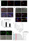
Grb4 is a key mediator to transduce EB3 reverse signals involved in axon pruning. a, Co-expression of Grb4 and EB3 in hippocampal granule cell neurons. b and c, Expression of dominant-negative Grb4-SH3Mut-DsRed (W38K, W148K and W234K) but not WT Grb4-DsRed (red fluorescence) in hippocampal neurons blocks axon pruning in live cells initiated by co-culture with CellTracker labeled Cos-1 cells (blue fluorescence) for 48 h (n = 11–16 neurons per group, * P < 0.0001). d, Expression of an EB3-Grb4-SH3 chimeric fusion protein in EB3−/− hippocampal neurons leads to axon shortening without addition of Cos-1 cells in live transfected neurons labeled with f-EGFP (upper panels) or in fixed cultures labeled with the indicated antibodies (lower panels). The EB3-Grb4-SH3Mut has no effect on axon shortening in EB3−/− neurons either with or without co-culture with Cos-1 cells. Arrowheads indicate axons and arrows indicate neuronal cell bodies. e, Quantification of neurite length in transfected EB3−/− neurons in the absence of Cos-1 co-culture (n = 8–10 neurons per group, * P < 0.0001). f, Expression of the EB3-Grb4-SH3 chimeric fusion protein in live differentiated NG108 cells is sufficient to mediate neurite shortening and rounding as indicated by co-expressed f-EGFP. g, Various EB3-Grb4-SH3 chimeric fusion proteins with indicated SH3 domain point mutations were scored for neurite length in live transfected NG108 cells. Quantification indicates the second SH3 domain is essential and sufficient to induce neurite shortening (n = 70–104 transfected cells per group). Mean ± s.e.m. Scale bars: 20 μm.
Figure 6
Figure 6
EB3 reverse signaling requires PAK and Dock180. a and b, Expression of dominant-negative Pak1DN-DsRed (indicated by red fluorescence) blocks neurite shortening induced by EB3-Grb4-SH3 (indicated by green f-EGFP fluorescence) in live NG108 cells (n = 45–89 transfected cells per group, *P < 0.001). c and d, Expression of Pak1DN-DsRed (red fluorescence) blocks axon pruning initiated by co-culture of primary hippocampal neurons with CellTracker labeled Cos-1 cells (blue) for 48 h in live cell assays (n = 11–13 transfected neurons per group, * P < 0.0001). e, Co-expression of Dock180 and EB3 in hippocampal granule cell neurons. f and g, Expression of dominant-negative Dock180-ISP (indicated by red immunofluorescence with anti-Flag) blocks axon pruning initiated by co-culture of primary hippocampal neurons with CellTracker labeled Cos-1 cells (blue) for 48 h (n = 10–14 transfected neurons per group, *P < 0.0001). Mean ± s.e.m. Scale bars: 20 μm. h, Grb4 and Dock180 co-immunoprecipitate with EB3-WT stably expressed in NG108 cells, but not with EB3-3F following 30 min EphB2-Fc stimulation of reverse signaling.
Figure 7
Figure 7
Activation of EB3 reverse signaling leads to increased Rac and Cdc42 GTP levels. a and b, Expression of a dominant-negative Rac1, mCherry-Rac1-N17, blocks axon pruning initiated by co-culture of primary hippocampal neurons with CellTracker labeled Cos-1 cells (blue) for 48 h in live cell assays (n = 13–27 transfected neurons per group, *P < 0.0001). Mean ± s.e.m. Scale bars: 20 μm. c, GTP-bound Rac1 and Cdc42 were precipitated by GST-PDB fusion protein pull downs from EB3-WT expressing NG108 cells following a time course of EphB2-Fc stimulation of reverse signaling. The immunoblots show a biphasic activation of Rac1 and Cdc42 in response to EphB2-Fc. Arrow indicates increased EB3 tyrosine phosphorylation. d, GTP-bound Rac1 and Cdc42 were precipitated by GST-PDB fusion protein pull downs from EB3-WT and EB3-3F expressing NG108 cells following 30 min EphB2-Fc stimulation of reverse signaling. The immunoblots show that Rac1 and Cdc42 are activated by EphB2-Fc stimulation in EB3-WT cells but not in EB3-3F cells.

References

    1. Patel BN, Van Vactor DL. Axon guidance: the cytoplasmic tail. Curr Opin Cell Biol. 2002;14:221–229. - PubMed
    1. Guan KL, Rao Y. Signalling mechanisms mediating neuronal responses to guidance cues. Nat Rev Neurosci. 2003;4:941–956. - PubMed
    1. Huber AB, Kolodkin AL, Ginty DD, Cloutier JF. Signaling at the growth cone: ligand-receptor complexes and the control of axon growth and guidance. Annu Rev Neurosci. 2003;26:509–563. - PubMed
    1. Luo L, O'Leary DD. Axon retraction and degeneration in development and disease. Annu Rev Neurosci. 2005;28:127–156. - PubMed
    1. Low LK, Cheng HJ. Axon pruning: an essential step underlying the developmental plasticity of neuronal connections. Philos Trans R Soc Lond B Biol Sci. 2006;361:1531–1544. - PMC - PubMed

Publication types

MeSH terms