. 2009 Feb;16(2):168-75.
doi: 10.1038/nsmb.1549. Epub 2009 Feb 1.

Helix movement is coupled to displacement of the second extracellular loop in rhodopsin activation

Affiliations

Helix movement is coupled to displacement of the second extracellular loop in rhodopsin activation

Shivani Ahuja et al. Nat Struct Mol Biol. 2009 Feb.

Abstract

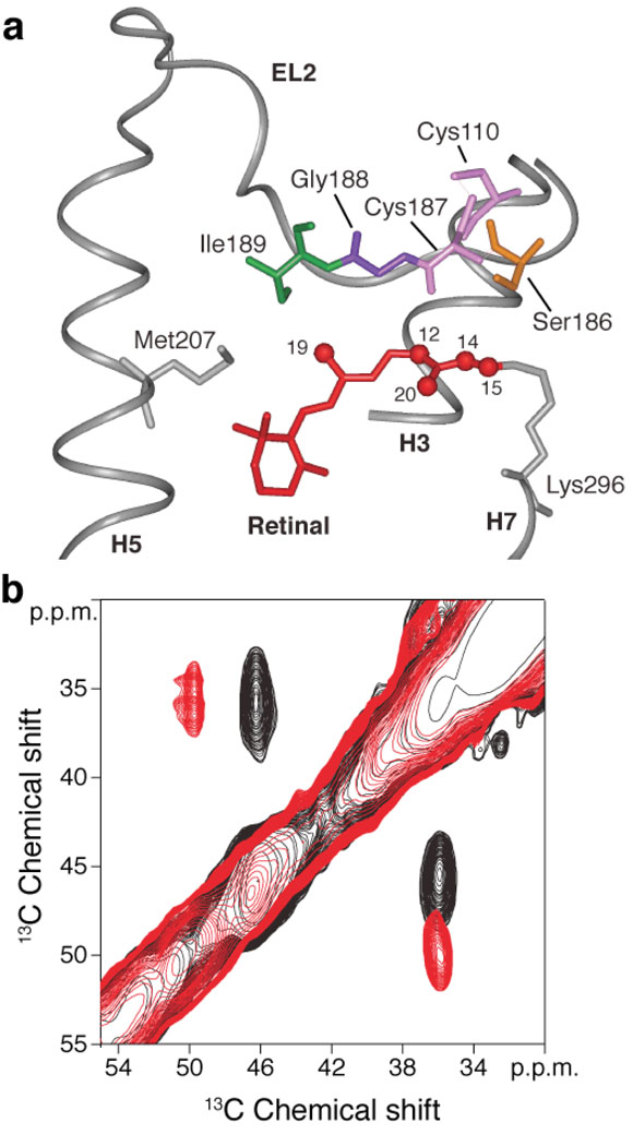
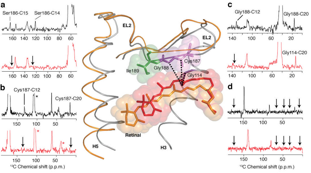
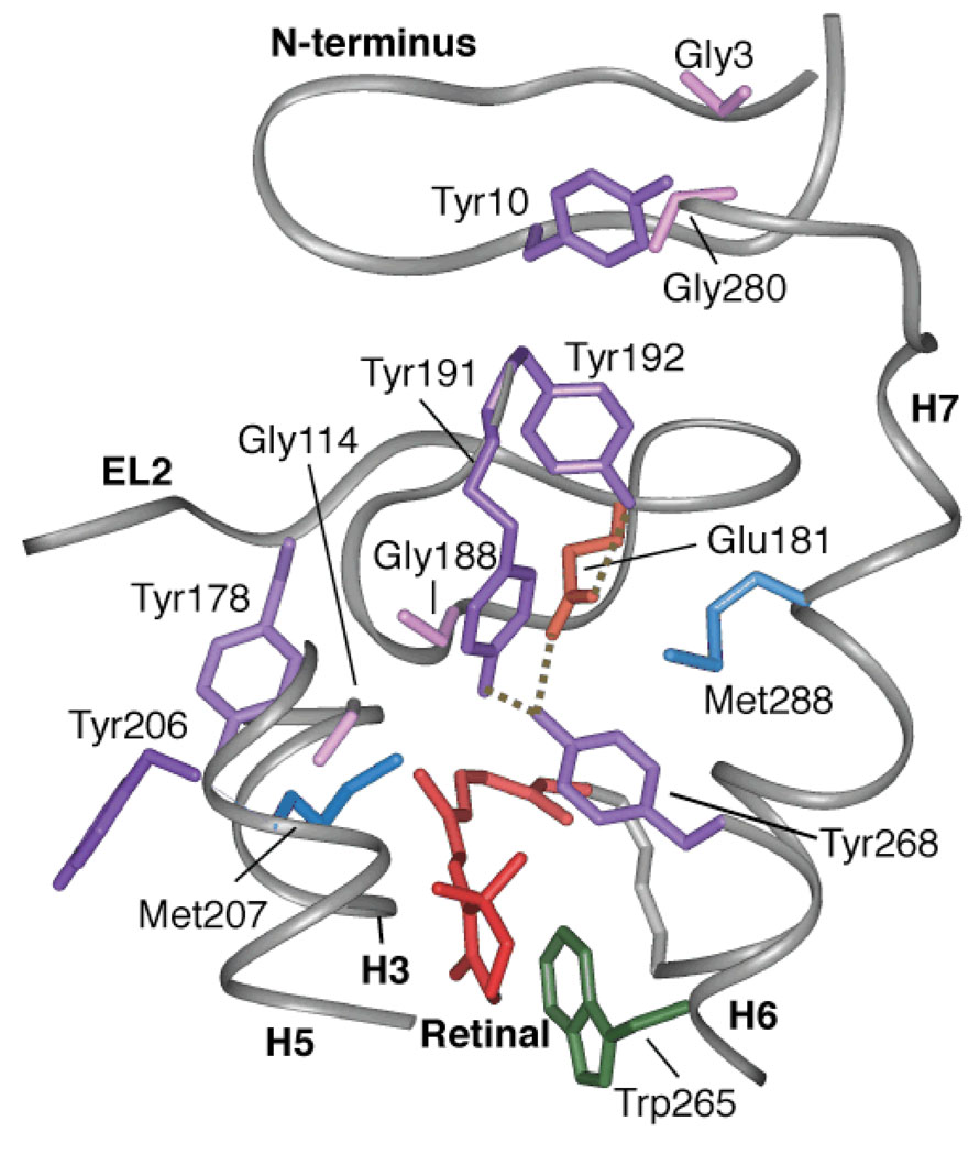
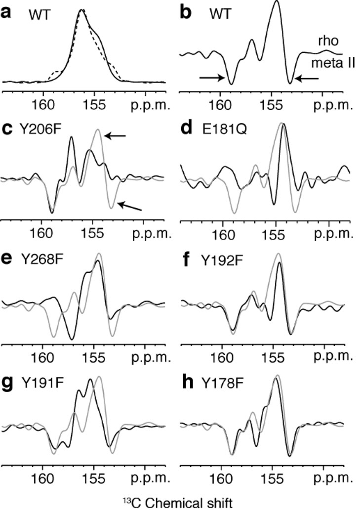
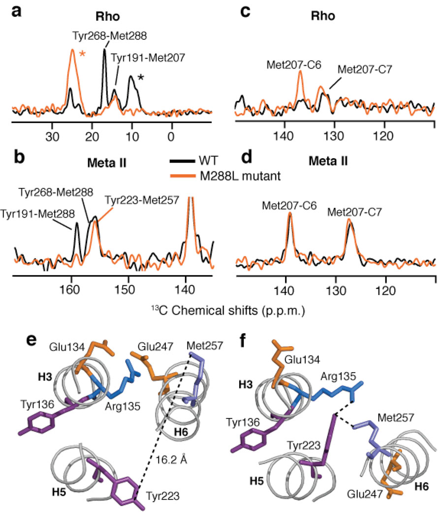
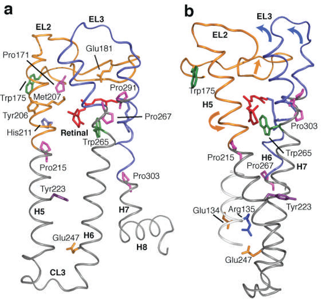
The second extracellular loop (EL2) of rhodopsin forms a cap over the binding site of its photoreactive 11-cis retinylidene chromophore. A crucial question has been whether EL2 forms a reversible gate that opens upon activation or acts as a rigid barrier. Distance measurements using solid-state (13)C NMR spectroscopy between the retinal chromophore and the beta4 strand of EL2 show that the loop is displaced from the retinal binding site upon activation, and there is a rearrangement in the hydrogen-bonding networks connecting EL2 with the extracellular ends of transmembrane helices H4, H5 and H6. NMR measurements further reveal that structural changes in EL2 are coupled to the motion of helix H5 and breaking of the ionic lock that regulates activation. These results provide a comprehensive view of how retinal isomerization triggers helix motion and activation in this prototypical G protein-coupled receptor.


Figures

Figure 1
Figure 1
Structural changes involving the conserved Cys110 - Cys187 disulfide link on activation of rhodopsin. (a) View of the β4 strand of EL2 from the rhodopsin crystal structure highlighting the interactions of Ile189, Gly188, Cys187 and Ser186 with the polyene chain of the retinal. Cys110 on the extracellular end of H3 forms a conserved disulfide link with Cys187 in β4. (b) A region from the 2D DARR NMR spectrum of rhodopsin selectively labeled with 13Cβ-cysteine. The figure highlights the crosspeak between Cys187 (46.8 ppm) and Cys110 (36.4 ppm) in rhodopsin (black). On conversion to meta II (red) there is a distinct shift in the crosspeak to 50.1 ppm for Cys187. The 13Cβ chemical shift of Cys110 at ~36 ppm does not change appreciably between rhodopsin and meta II. The eight reduced cysteines in rhodopsin are observed as a broad resonance at ~25 ppm (not shown).
Figure 2
Figure 2
2D 13C DARR NMR spectra of retinal – EL2 interactions. Rows are shown from the 2D 13C DARR NMR spectra of rhodopsin (black) and meta II (red). (a) Rhodopsin labeled with 13Cβ-serine and 13C14,15 retinal. Crosspeaks are observed between Ser186 (63.3 ppm) and the 13C14 and 13C15 resonances in dark rhodopsin, which are lost (arrows) in meta II. (b) Rhodopsin labeled with 13C1-cysteine and 13C12,20 retinal. Crosspeaks are observed between Cys187 (170.8 ppm) and the 13C12 and 13C20 resonances in dark rhodopsin, which are lost (arrows) in meta II. (c) Rhodopsin labeled with 13Cα-glycine and 13C12,20 retinal. Crosspeaks are observed between Gly188 (42.0 ppm) and the 13C12 and 13C20 resonances in dark rhodopsin, which are lost (arrows) in meta II. However, a new Gly-C20 contact is observed in meta II, which is assigned to Gly114 (see text). (d) Rhodopsin labeled with U-13C6-isoleucine and 13C9 retinal. No contacts were observed between Ile189 and C9 on the polyene chain of the retinal in either rhodopsin (black arrows) or meta II (red arrows). The structure of EL2 in rhodopsin is shown (center) indicating the contacts observed between the C20 methyl group and Cys187, Gly188 and Gly114 in rhodopsin. In order to illustrate the displacement of EL2 needed to satisfy the NMR constraints, we have superimposed the rhodopsin crystal structure (grey) with the meta II model (orange) obtained from MD simulations guided by our experimentally determined retinal-protein contacts. Asterisks correspond to MAS sidebands.
Figure 3
Figure 3
A view of the extracellular side of the rhodopsin from the crystal structure. The figure highlights the relative position of six tyrosines: Tyr10, Tyr178, Tyr191, Tyr192, Tyr206 and Tyr268. Of these tyrosines, Tyr191, Tyr192 and Tyr268 are involved in the H-bonding network with Glu181. Tyr268 and Tyr191 are also in close contact with Met288 on H7. Tyr206 on H5 is involved in a second H-bonding network with His211 (H5), Glu122 (H3), Trp126 (H3) and Ala166 (H4) (not shown). Additionally, the figure shows Tyr-Gly interactions on the extracellular side of rhodopsin between Gly188-Tyr268, Gly3-Tyr10-Gly280 and Gly114-Tyr178.
Figure 4
Figure 4
One dimensional (1D) 13C CP-MAS spectra of rhodopsin and meta II labeled with 13Cζ-tyrosine. (a) Overlap of the 13C 1D CP-MAS spectra of the 13Cζ-tyrosine resonance in rhodopsin (black) and meta II (dashed black). Difference spectra for wild-type rhodopsin (b) and several rhodopsin mutants, Y206F (c), E181Q (d), Y268F (e), Y192F (f), Y191F (g), Y178F (h). The difference spectrum in gray corresponds to the wild-type protein.
Figure 5
Figure 5
2D DARR NMR of Tyr(Cζ) - Met(Cε) contacts in rhodopsin and the M288L rhodopsin mutant. (a) Rows through the 13Cζ-Tyr diagonal resonance from 2D DARR NMR spectra of rhodopsin (black) and the M288L rhodopsin mutant (orange) labeled with 13Cζ-tyrosine and 13Cε-methionine. (b) Rows through the 13Cε-Met diagonal resonance from 2D DARR NMR spectra of meta II (black) and the M288L rhodopsin mutant (orange) following conversion to meta II. (c) Rows through the 13Cε-methionine diagonal resonance of rhodopsin (black) and the M288L rhodopsin mutant (orange) showing the crosspeaks to the retinal 13C6 and 13C7 resonances. (d) Same as in (c) following conversion to meta II. In the M288L mutant of rhodopsin, we observe a contact between Met207 and C6 that is not present in wild type rhodopsin. This change in the Met207-retinal contact in the M288L mutant of rhodopsin can be interpreted as either a change in the position of the retinal or in the position of Met207 on H5 upon mutation of Met288 (H7) to a leucine. Upon activation, the Met207 – retinal interactions in the M288L mutant are identical to those in wild-type meta II. (e) A view of the ionic lock between Arg135 and Glu247 from the crystal structure of rhodopsin. The Tyr223-Met257 distance is well beyond the range of the DARR NMR experiment. (f) Structure of the ionic lock from the recent crystal structure of opsin, showing the close proximity between Tyr223 and Met257. Asterisks correspond to MAS sidebands.
Figure 6
Figure 6
Crystal structure of rhodopsin highlighting EL2 and H5. (a) Retinal isomerization within the tightly packed binding site results in steric contacts between the β-ionone ring and H5, and between the retinal C19 and C20 methyl groups and EL2. These interactions trigger the simultaneous displacement of EL2 and H5. Motion of the β-ionone ring is also coupled to the motion of Trp265. Trp265 is packed against the β-ionone ring and C20 of the retinal, as well as Gly121 on H3 and Ala295 on H7. Movement of the Trp265 sidechain away from these critical contacts allows helices H6 and H7 to shift into active conformations. The coupled motions of helices H5–H7, in turn, are coupled to the rearrangement of electrostatic interactions involving the conserved ERY sequence at the cytoplasmic end of H3, exposing the G protein binding site on the cytoplasmic surface of the protein. (b) View of the rhodopsin crystal structure highlighting the interaction between EL2 and EL3 on the extracellular side of the receptor, and the positions of Tyr223 and the conserved Glu135-Arg135-Tyr136 sequence on the intracellular side of the receptor.

Similar articles

Cited by

References

    1. Samson M, et al. The second extracellular loop of CCR5 is the major determinant of ligand specificity. J. Biol. Chem. 1997;272:24934–24941. - PubMed
    1. Shi L, Javitch JA. The second extracellular loop of the dopamine D-2 receptor lines the binding-site crevice. Proc. Natl. Acad. Sci. U.S.A. 2004;101:440–445. - PMC - PubMed
    1. Klco JM, Wiegand CB, Narzinski K, Baranski TJ. Essential role for the second extracellular loop in C5a receptor activation. Nat. Struct. Mol. Biol. 2005;12:320–326. - PubMed
    1. Scarselli M, Li B, Kim SK, Wess J. Multiple residues in the second extracellular loop are critical for M-3 muscarinic acetylcholine receptor activation. J. Biol. Chem. 2007;282:7385–7396. - PubMed
    1. Palczewski K, et al. Crystal structure of rhodopsin: A G protein-coupled receptor. Science. 2000;289:739–745. - PubMed

Publication types