. 2009 Mar;10(3):257-65.
doi: 10.1038/ni.1697. Epub 2009 Feb 1.

The peptidyl-prolyl isomerase Pin1 facilitates cytokine-induced survival of eosinophils by suppressing Bax activation

Affiliations

The peptidyl-prolyl isomerase Pin1 facilitates cytokine-induced survival of eosinophils by suppressing Bax activation

Zhong-Jian Shen et al. Nat Immunol. 2009 Mar.

Abstract

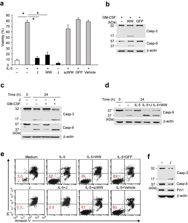
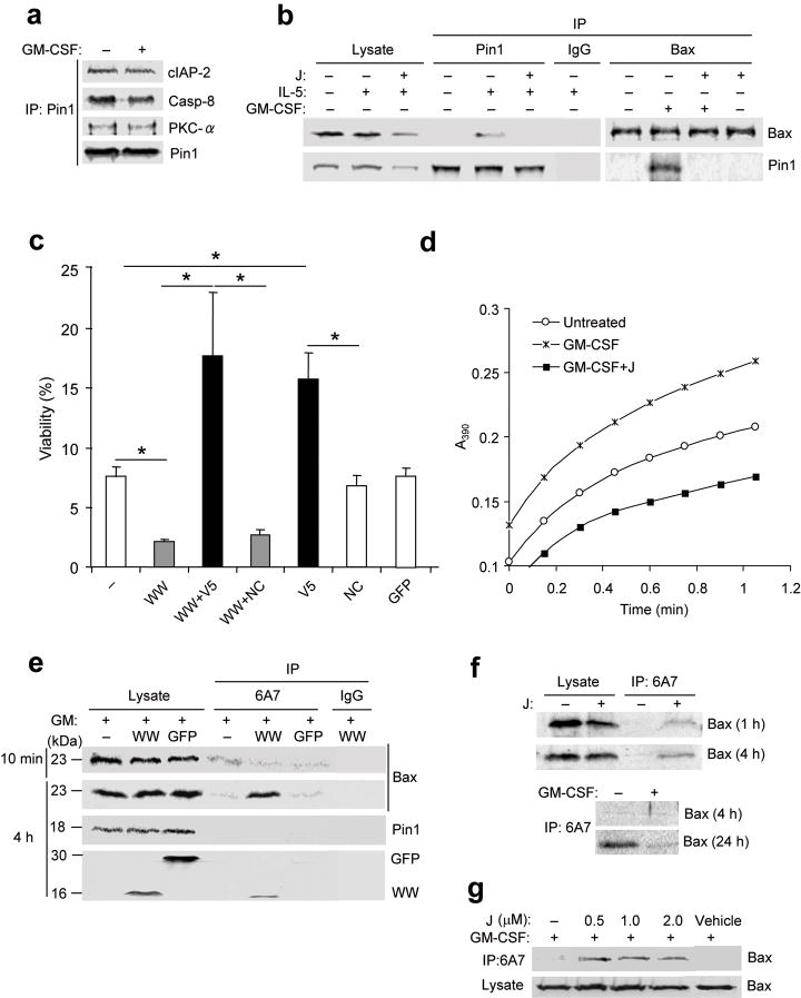
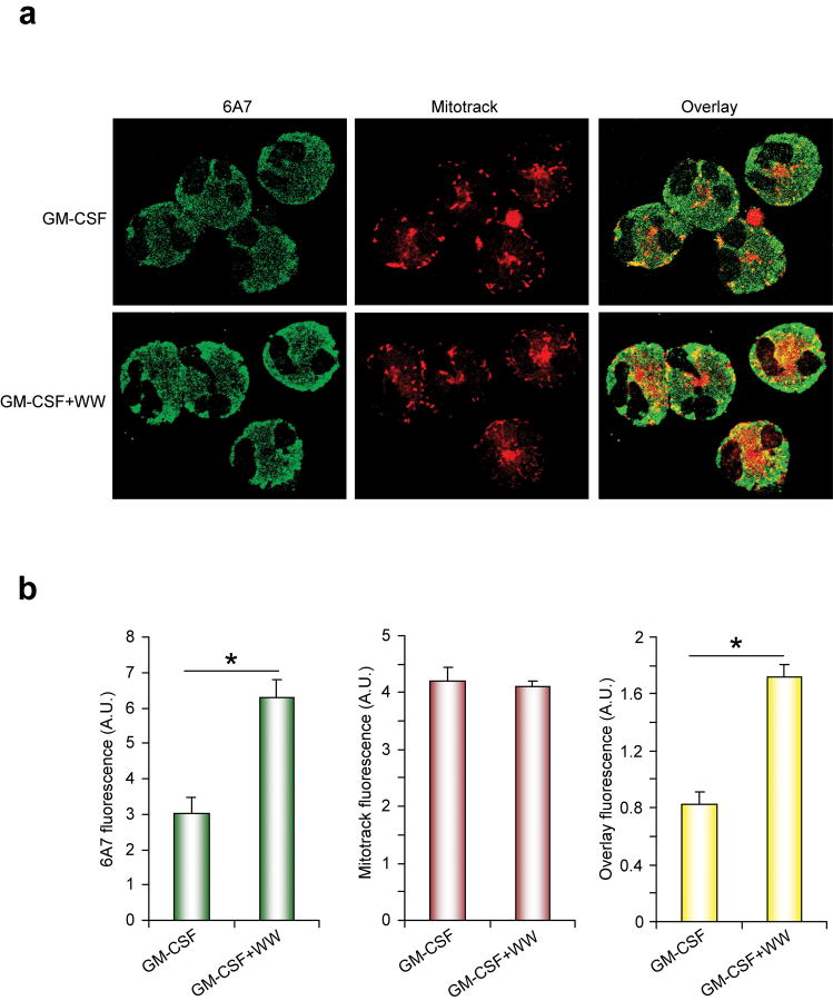
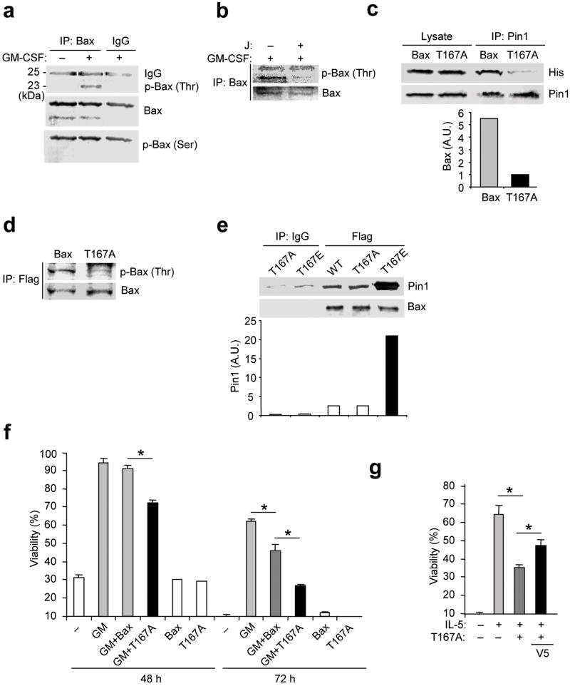
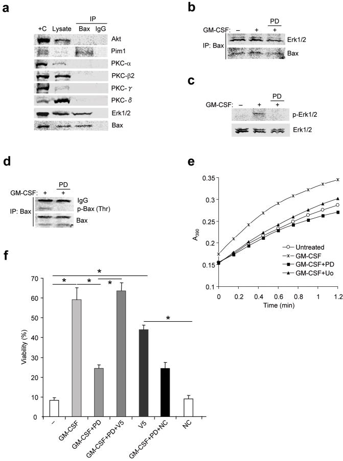
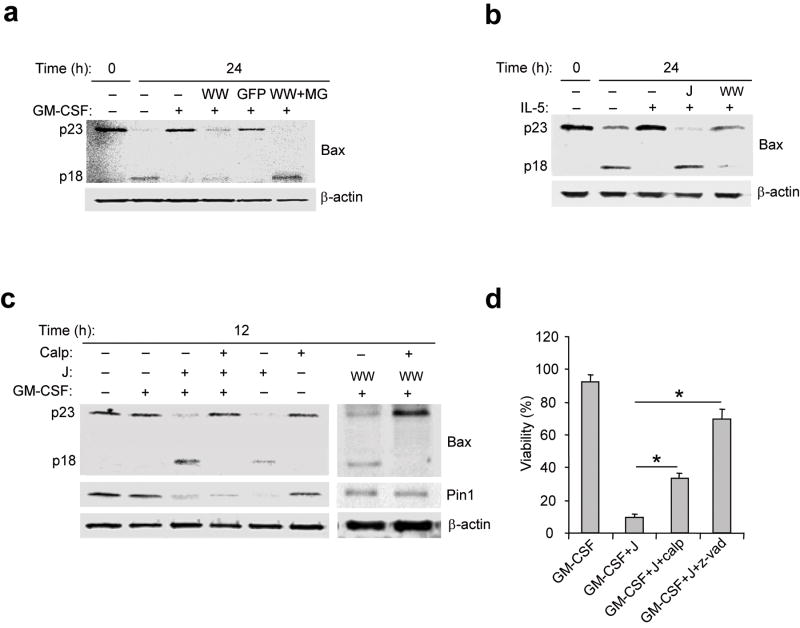
The mechanisms by which cytokine signals prevent the activation and mitochondrial targeting of the proapoptotic protein Bax are unclear. Here we show, using primary human eosinophils, that in the absence of the prosurvival cytokines granulocyte-macrophage colony-stimulating factor and interleukin 5, Bax spontaneously underwent activation and initiated mitochondrial disruption. Inhibition of Bax resulted in less eosinophil apoptosis, even in the absence of cytokines. Granulocyte-macrophage colony-stimulating factor induced activation of the kinase Erk1/2, which phosphorylated Thr167 of Bax; this facilitated new interaction of Bax with the prolyl isomerase Pin1. Blockade of Pin1 led to cleavage and mitochondrial translocation of Bax and caspase activation, regardless of the presence of cytokines. Our findings indicate that Pin1 is a key mediator of prosurvival signaling and is a regulator of Bax function.


Conflict of interest statement

Competing interests statement. The authors declare no competing financial interests.

Figures

Figure 1
Figure 1. Pin1 blockade suppresses cytokine-induced survival
(a) Viability of purified eosinophils left untreated (–) or incubated for 3 days with IL-5 (200 pM) and/or with juglone (J; 1 μM), TAT-WW-Pin1 (WW; 300 nM), scrambled WW peptide (scWW; 300 nM), TAT-GFP (GFP; 300 nM), or vehicle (ethanol). Cell viability was assessed in triplicate cultures from 3 different donors by trypan blue exclusion, and expressed as a percentage of the viability at time 0. *, P < 0.05 by Student’s t-test in a two-tailed analysis. (b-d, f) Immunoblots of total Eos lysates. Anti-caspase 3 detects the “pro-form” (p32) and the cleaved, active form (p17). Anti-caspase 9 detects the “pro-form” (p57) and the cleaved, active form (p37). (b) Eosinophils were incubated for 24 h with GM-CSF (100 pg/ml) alone or together with TAT-WW-Pin1 or TAT GFP. (c,d) Cells were left untreated (–) and lysed immediately (0 h) or incubated for 24 h in culture alone (–), or with GM-CSF or IL-5 alone or together with juglone or TAT-WW-Pin1. (e) Representative flow cytometric analysis of eosinophil viability 72 h after exposure to the indicated treatment. Eosinophils were stained with PI and annexin V. Percentage of viable cells is shown in the lower left quadrant. (f) Purified eosinophils from BAL fluid were obtained from subjects after allergen challenge. Cells were left untreated (–) or incubated with juglone for 24 h prior to immunoblot with antibodies specific for the indicated proteins. Immunoblots are representative of results from at least 3 different donors.
Figure 2
Figure 2. Pin1 interacts with Bax and regulates its activation
(a-b) Eosinophils were left untreated (–) or incubated for 4 h with cytokine alone or with juglone (J). Cells were lysed in zwitterionic detergent (CHAPS) buffer and the lysates (10% used for immunoblot) were pre-cleared with nonimmune IgG and immunoprecipitated (IP) followed by immunoblot as shown. (c) Eosinophils were left untreated (–) or incubated for 3 days with TAT-WW-Pin1 (WW) or TAT-GFP (GFP), with or without V5 (500 μM) or a negative control peptide (NC). Cell viability of triplicate cultures from 3 different donors was determined by trypan blue exclusion, and expressed as a percentage of the viability at time 0. *, P < 0.05 by Student’s t-test in a two-tailed analysis. (d) Pin1 isomerase assay of cytoplasmic lysates from eosinophils left untreated or after treatment for 10 min with GM-CSF alone or together with juglone. (e) Eosinophils were treated with GM-CSF alone or together with His-TAT-WW-Pin1 or His-TAT-GFP for 10 min or 4 h. Lysates were subjected to immunoprecipitation with the antibody 6A7 or nonimmune IgG, followed by immunoblot. Anti-His was used to detect His-GFP-TAT and His-TAT-WW-Pin1. (f) Cells were incubated without (top) or with GM-CSF (bottom), with or without juglone for the times shown. Lysates were subjected to immunoprecipitation with 6A7 and immunoblot with anti-Bax. (g) Cells were treated with GM-CSF alone or together with juglone at the indicated dose. Cell lysates were immunoprecipitated with 6A7 followed by immunoblot with anti-Bax. Immunoblots and isomerase assay are representative of at least 3 experiments with different donors.
Figure 3
Figure 3. Pin1 blockade causes mitochondrial translocation of Bax
(a) Eosinophils were incubated for 4 h with GM-CSF alone or with TAT-WW-Pin1. Mitotracker (red; 75 nM) was added to culture medium 45 min before harvest. Cytospins were prepared and stained with 6A7 antibody (green). (b) The density of fluorescence staining for >50 cells was quantified as described in Methods. *, P < 0.05 by Student’s t-test in a two-tailed analysis. Results are representative of at least 3 experiments with different donors.
Figure 4
Figure 4. Phosphorylation of Bax Thr167 facilitates cell survival
(a) Eosinophils were left untreated (–) or incubated for 4 h with GM-CSF. Cells were lysed in CHAPS buffer and immunoprecipitated (IP) with anti-Bax or nonimmune IgG, followed by immunoblot with anti-p-Thr, anti-p-Ser and anti-Bax. (b) Eosinophils were treated with GM-CSF alone or together with juglone as in (a). Cell lysates were immunoprecipitated with anti-Bax prior to immunoblot with anti-p-Thr and anti-Bax. (c) Eosinophils were incubated with His-tagged TAT-WT-Bax (Bax) or TAT-T167A-Bax (T167A) for 10 min in the presence of GM-CSF. Cell lysates were pre-cleared and immunoprecipitated with anti-Pin1 followed by immunoblot with anti-His and anti-Pin1. Top, representative immunoblot. Bottom, the density of Bax and T167A bands, normalized to the Pin1 bands. (d) Eosinophils were incubated with Flag-tagged TAT-WT-Bax or TAT-T167A-Bax. After treatment with GM-CSF, cell lysates were immunoprecipitated with anti-Flag followed by immunoblot with anti-p-Thr and anti-Bax. (e) WT, T167A and T167E Bax proteins were incubated in vitro with GST-Pin1 in CHAPS buffer prior to immunoprecipitation with anti-Flag or nonimmune IgG followed by immunoblot with anti-Pin1 or anti-Bax. Top, representative immunoblot. Bottom, the density of Pin1 bands, normalized to the Bax bands. (f) Eosinophils were incubated with or without Flag-tagged TAT-WT-Bax or TAT-T167A-Bax in the presence or absence of GM-CSF (50 pg/ml). Cell viability was determined after 48 and 72 h. (g) Cells were incubated with or without IL-5 (100 pM) alone or together with TAT-Bax-T167A or V5. Cell viability was determined after 72 h). *, P < 0.05 Student’s t-test in a two-tailed analysis. Immunoblots are representative of at least 3 experiments with different donors.
Figure 5
Figure 5. Bax associates with and is phosphorylated by Erk1/2
(a) Eosinophils were treated with GM-CSF for 1 h. Cells were lysed in CHAPS buffer and pre-cleared with nonimmune IgG prior to immunoprecipitation with anti-Bax (total) or nonimmune IgG followed by immunoblot as shown. +C: positive control (mouse brain lysate), ; L: 10% of initial lysate. (b-e) Eosinophils were pre-incubated with the MEK1 inhibitors PD98059 (PD; 50 μM) or U0126 (Uo; 10 μM) for 30 min before stimulation with GM-CSF for 1 h. (b-d) Cells were lysed in CHAPS buffer and the lysates were pre-cleared with nonimmune IgG and immunoprecipitated with anti-Bax followed by immunoblot (b), or lysates were immunoblotted with antibodies specific for p-Erk1/2 and total Erk1/2 (c) or anti-p-Thr and anti-Bax (d). (e) PPIase activity in lysates was measured (See Methods). Uo: U0126 (10 μM). (f) Cells were pre-incubated with PD98059 for 1 h before incubation with GM-CSF (50 pg/ml) with or without the V5 Bax inhibitory peptide (V5, 500 μM) or a control peptide (NC) for 72 h. Cell viability was determined by trypan blue exclusion, and viability was expressed as a percentage of viability at time 0. *, P < 0.05 by Student’s t-test in a two-tailed analysis. Immunoblot and isomerase assays are representative results from at least 3 different donors. Cell viability was assessed in triplicate cultures from 3 different donors.
Figure 6
Figure 6. Bax activity and cleavage are modified by calpain inhibitors and cytokine signaling
(a-c) Immunoblots of total eosinophil lysates. Anti-Bax (clone 3) detects the “pro-form” (p23) and the cleaved form (p18). (a) Eosinophils were left untreated (–) and immediately lysed (0) or incubated for 24 h with GM-CSF alone or together with TAT-WW-Pin1 (WW), TAT-GFP (GFP) or TAT-WW-Pin1 and MG132 (MG; 40 μM). (b) Freshly purified eosinophils were immediately lysed (0) or left untreated for 24 h or incubated with IL-5 alone or together with juglone (J) or TAT-WW-Pin1. (c) Eosinophils were left untreated or incubated with GM-CSF alone or together with juglone or TAT-WW or calpeptin (Calp, 20 μM). (d) Eosinophils were treated with GM-CSF alone or with juglone or calpeptin or with the broad spectrum caspase inhibitor z-Vad-fmk (z-vad; 50 μM). Viability was determined after 72 h incubation. Cell viability was determined by trypan blue exclusion, and viability was expressed as a percentage of viability at time 0. *, P < 0.05 Student’s t-test in a two-tailed analysis. Immunoblots are representative of at least 3 experiments with different donors while cell viability was assessed in triplicate cultures from 3 different donors.

Similar articles

Cited by

References

    1. Brightling CE, et al. Comparison of airway immunopathology of eosinophilic bronchitis and asthma. Thorax. 2003;58:528–532. - PMC - PubMed
    1. Shen ZJ, et al. Pin1 regulates TGF-beta1 production by activated human and murine eosinophils and contributes to allergic lung fibrosis. J Clin Invest. 2008;118:479–490. - PMC - PubMed
    1. Gibson PG, Saltos N, Borgas T. Airway mast cells and eosinophils correlate with clinical severity and airway hyperresponsiveness in corticosteroid-treated asthma. J Allergy Clin Immunol. 2000;105:752–759. - PubMed
    1. Flood-Page P, et al. Anti-IL-5 treatment reduces deposition of ECM proteins in the bronchial subepithelial basement membrane of mild atopic asthmatics. J Clin Invest. 2003;112:1029–1036. - PMC - PubMed
    1. Humbles AA, et al. A critical role for eosinophils in allergic airways remodeling. Science. 2004;305:1776–1779. - PubMed

Publication types

MeSH terms

Substances