. 2009 Mar;10(3):314-24.
doi: 10.1038/ni.1698. Epub 2009 Feb 1.

The interleukin 23 receptor is essential for the terminal differentiation of interleukin 17-producing effector T helper cells in vivo

Affiliations

The interleukin 23 receptor is essential for the terminal differentiation of interleukin 17-producing effector T helper cells in vivo

Mandy J McGeachy et al. Nat Immunol. 2009 Mar.

Abstract

Interleukin 23 (IL-23) is required for autoimmune inflammation mediated by IL-17-producing helper T cells (T(H)-17 cells) and has been linked to many human immune disorders. Here we restricted deficiency in the IL-23 receptor to defined cell populations in vivo to investigate the requirement for IL-23 signaling in the development and function of T(H)-17 cells in autoimmunity, inflammation and infection. In the absence of IL-23, T(H)-17 development was stalled at the early activation stage. T(H)-17 cells failed to downregulate IL-2 and also failed to maintain IL-17 production or upregulate expression of the IL-7 receptor alpha-chain. These defects were associated with less proliferation; consequently, fewer effector T(H)-17 cells were produced in the lymph nodes and hence available to emigrate to the bloodstream and tissues.


Figures
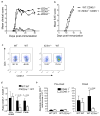
Figure 1
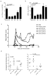
Figure 1. Il23ra−/− CD4+ T cells do not accumulate in the inflamed CNS
(a) Clinical scores of Il23ra−/−, Il23ra+/− and Il23ra+/+ mice after EAE induction. (b) Clinical scores of mixed bone marrow chimeras following EAE induction. Donor bone marrow genotype is indicated. (c) Flow cytometry of CD4+ cells obtained from the CNS of mixed bone marrow chimeras 11 days post-EAE induction. Genotype of each population is indicated. (d) Proportion of wild-type CD45.1+ or Il23ra−/− CD45.1− CD4+ T cells in the CNS of mixed bone marrow chimeras at indicated time points; data is expressed as mean ratio ± s.d. of CD45.1− to CD45.1+ CD4+ T (wild-type) cells. (e) Absolute numbers of CD4+ T cells in the CNS on day 9 (pre-onset) and day 11 (onset of clinical signs) of EAE. Data shown are each representative of three experiments with 5 mice per group.
Figure 2
Figure 2. Il23ra−/− T cells have reduced IL-17 production after T cell priming
(a) CD4+ IL-17 production after 24 hour stimulation of CNS cells with MOG(35–55) in the presence or absence of IL-23, CNS cells isolated from mixed bone marrow chimeras 11 days post-EAE induction. (b) CD4+ IFN-γ production after 24 hour stimulation of CNS cells with MOG(35–55) or medium only control, CNS cells isolated from mixed bone marrow chimeras 11 days post-EAE induction. (c) Supernatant IL-17 concentrations after stimulation of CNS mononuclear cells as described in a. (d) Mean fluorescence intensity of IL-17+ wild-type CD4+ CNS cells stimulated as in a. (e) Supernatant IFN-γ concentrations after stimulation of CNS mononuclear cells as in a. (f) Draining lymph node CD4+ T cell IL-17 production in response to 24 hour stimulation with MOG(35–55) in the presence or absence of IL-23, 11 days post-EAE induction. Data shown are from 5 mice per group, representative of at least 3 experiments with similar results.
Figure 3
Figure 3. Early TH-17 activation is normal but effector responses are impaired in Il23ra −/−
(a) Frequency of wild-type or Il23ra−/− CD45.1+CD4+OTII T cells, expressed as percentage of total CD4+ T cells (mean ± s.d), following transfer into wild-type recipients and immunization with OVA(323–339). (b) Delayed type hypersensitivity reaction in mice after transfer of wild-type or Il23ra−/− OTII cells. The increase in foot thickness over the contralateral saline-challenged foot is shown. (c) Time course of Intracellular IL-17 in OTII cells from dLN assessed by flow cytometry after ex vivo stimulation with PMA + ionomycin on indicated days post-immunization. (d) Intracellular Foxp3 expression in OTII cells assessed on days 5 and 7 post-immunization (e) Flow cytometry for phosphorylated STAT3 in OTII cells after 15 minute stimulation with indicated cytokines 4 days post-immunization. (f) Flow cytometry of Cre-deleted Stat3flox/flox CD4+ cells following second stimulation with anti-CD3 in the presence of IL-23. IL-17 is shown as percentage of GFP+ and GFP− cells on each plot. All data shown are representative of at least 3 independent experiments having 4 to 5 mice per group.
Figure 4
Figure 4. Full differentiation of effector TH-17 cells requires IL-23R in vivo.
(a–e) Analysis of phenotype of wild-type or Il23ra−/− CD45.1+CD4+OTII T cells after transfer into wild-type recipients and immunization with OVA(323–339). (a) Flow cytometry for cell surface expression of CD44, CCR7 and CD62L on wild-type and Il23ra−/− OTII T cells (shaded histograms) overlaid with total CD4+ cells for comparison (clear histograms) at day 7 post-immunization. b) Surface IL-7Rα expression on wild-type (shaded histogram) and Il23ra−/− (clear histogram) OTII cells from dLN. c) RT-PCR analysis of IL-7Rα expression in OTII+ cells sorted by flow cytometry from dLN; values are normalized to ubiquitin expression. As a control, total OTII–CD4+ cells were isolated at the same time from recipients of the indicated OTII cells. Data shown are mean ± s.d. of two groups of samples each containing 5 mice per condition. d) Surface CD27 expression on OTII cells in dLN and blood. e) Frequency of wild-type and Il23ra−/− OTII cells (as proportion of total CD4+ cells) in blood at indicated time points post immunization. Data shown are representative of 3 experiments with similar results.
Figure 5
Figure 5. IL-23 promotes effector cytokine profile of TH-17
(a–d) Intracellular cytokine analysis of wild-type or Il23ra−/− CD45.1+CD4+OTII T cells after transfer into wild-type recipients and immunization with OVA(323–339). (a) Intracellular staining of IL-17 and IL-2 in dLN OTII cells (b) Mean frequency of IL-17+IL-2− cells as proportion of CD4+OTII cells at indicated time points post-immunization. (c) Mean frequency of IL-2+IL-17− cells as proportion of CD4+OTII cells at indicated time points post-immunization. (d) Intracellular staining of IL-17 and IL-2 in blood OTII. Data shown are representative of 4 independent experiments with 4 mice per group.
Figure 6
Figure 6. IL-23R is not required for TH1 effector cell development
(a–b) Intracellular cytokine analysis of wild-type or Il23ra−/− CD45.1+CD4+OTII T cells after transfer into wild-type recipients and immunization with OVA(323–339). a) Frequency of IFN-γ+IL-17− cells as a proportion of CD4+OTII cells on indicated days post-immunization. b) Frequency of IFN-γ+IL-17+ cells as a proportion of CD4+ OTII cells on indicated days post-immunization. Data shown are representative of 3 independent experiments with 5 mice per group. (c–e) Wild-type and Il23ra−/− mice were infected with T. gondii and then followed for survival (c); frequency of IFN-γ+CD4+ cells following ex vivo stimulation of dLN cells from mice 9 days post-infection were also evaluated (d). (e) Intracellular flow cytometry of IFN-γ and IL-2 production by CD4+ cells following ex vivo stimulation of dLN cells from mice 9 days post-infection Data shown are representative of two independent experiments (5 mice per group) with similar results.
Figure 7
Figure 7. Timing of IL-23R requirement during TH-17 differentiation
(a,b) Anti-IL-23R was administered to recipients of wild-type or Il23ra−/− OTII cells at the indicated times postimmunization. Draining lymph nodes were harvested on day 10 for analysis of intracellular IL-17 and IL-2 (a) and frequency of OTII cells in blood was assessed (b). (c) Clinical score of SJL mice administered anti-IL-23R on days indicated after EAE induction. (d) Delayed type hypersensitivity response in footpads of p19-deficient recipients of wild-type OTII cells ten days postimmunization with OVA(323–339). Where indicated, IL-23 was administered with ovalbumin in the footpad at the time of challenge and mice also received IL-23 in the contralateral footpad. Data shown are each representative of at least two experiments with 5 mice per group.
Figure 8
Figure 8. Proliferation of IL-17+ cells is most affected by IL-23R deficiency
(a) Frequency of OTII+ cells expressing CCR6 in dLN on day 10 post-immunization. (b) S1p1 gene expression in OTII+ cells sorted by flow cytometry from dLN on day 7, values are normalized to ubiquitin. As a control, total OTII–CD4+ cells were isolated at the same time from recipients of the indicated OTII cells. (c) Proportion of IL-17+ cells that were positive for active caspase 3 was assessed by intracellular staining in OTII cells from dLN at indicated times post-immunization. (d) IL-21 expression in OTII+ cells sorted by flow cytometry from dLN on day 7; values are normalized to ubiquitin. (e.f) Analysis of BrdU incorporation by OTII cells 4 hour after in vivo administration of BrdU on indicated days post-immunization; expressed as proportion of IL-17+ (e) and IL-17− (f) OTII cells that were positive for BrdU. Data shown are representative of 2 independent experiments each with 4–5 mice/group, except (e,f) representative of 4 independent experiments with 4 mice per group.

Comment in

Similar articles

Cited by

References

    1. Oppmann B, et al. Novel p19 protein engages IL-12p40 to form a cytokine, IL-23, with biological activities similar as well as distinct from IL-12. Immunity. 2000;13:715–25. - PubMed
    1. Cua DJ, et al. Interleukin-23 rather than interleukin-12 is the critical cytokine for autoimmune inflammation of the brain. Nature. 2003;421:744–8. - PubMed
    1. McKenzie BS, Kastelein RA, Cua DJ. Understanding the IL-23–IL-17 immune pathway. Trends Immunol. 2006;27:17–23. - PubMed
    1. Langrish CL, et al. IL-23 drives a pathogenic T cell population that induces autoimmune inflammation. J Exp Med. 2005;201:233–40. - PMC - PubMed
    1. Murphy CA, et al. Divergent pro- and antiinflammatory roles for IL-23 and IL-12 in joint autoimmune inflammation. J Exp Med. 2003;198:1951–7. - PMC - PubMed

MeSH terms

LinkOut - more resources