. 2009 Mar 24;48(11):2538-49.
doi: 10.1021/bi802198q.

Isolation of mammalian 26S proteasomes and p97/VCP complexes using the ubiquitin-like domain from HHR23B reveals novel proteasome-associated proteins

Affiliations

Isolation of mammalian 26S proteasomes and p97/VCP complexes using the ubiquitin-like domain from HHR23B reveals novel proteasome-associated proteins

Henrike C Besche et al. Biochemistry. .

Abstract

Recent studies, mainly in yeast, have identified various cofactors that associate with the 26S proteasome and appear to influence its function. To identify these proteins in different cells and physiological states, we developed a method to gently and rapidly isolate 26S proteasomes and associated proteins without the need for genetic modifications of the proteasome. This method is based on the affinity of this complex for the ubiquitin-like (UBL) domain of hHR23B and elution with a competing polypeptide containing a ubiquitin-interacting motif. Associated with 26S proteasomes from rat muscle were a variety of known proteasome-interacting proteins, activators, and ubiquitin conjugates. In addition, we identified over 40 proteins not previously known to associate with the 26S proteasome, some of which were tightly associated with the proteasome in a substoichiometric fashion, e.g., the deubiquitinating enzymes USP5/isopeptidase T and USP7/HAUSP and the ubiquitin ligases ARF-BP1/HUWE1 and p600/UBR4. By altering buffer conditions, we also purified by this approach complexes of the ATPase p97/VCP associated with its adaptor proteins Ufd1-Npl4, p47, SAKS1, and FAF1, all of which contain ubiquitin-binding motifs. These complexes were isolated with ubiquitin conjugates bound and were not previously known to bind to the UBL domain of hHR23B. These various UBL-interacting proteins, dubbed the UBL interactome, represent a network of proteins that function together in ubiquitin-dependent proteolysis, and the UBL method offers many advantages for studies of the diversity, functions, and regulation of 26S proteasomes and p97 complexes under different conditions.


Figures
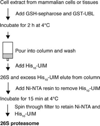
Figure 1
Figure 1
Flow chart of affinity method to isolate 26S proteasomes from mammalian cells and tissues. GST-UBL, recombinant fusion protein of glutathione S-transferase (GST) and the ubiquitin-like domain (UBL) of human HR23B; His10-UIM, recombinant protein containing the second ubiquitin-interacting motif of human S5a; GSH-Sepharose, glutathione-coupled resin with affinity for GST; Ni-NTA, nickel-charged nitrilotriacetic acid (NTA) agarose that binds to His tags.
Figure 2
Figure 2
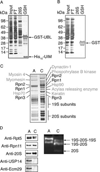
The UBL-affinity purification yields pure 26S proteasomes. (A) Muscle extract (2 g of rat skeletal muscle/10 mL of buffer) was cleared by 1 h 100000g ultracentrifugation (input) and incubated with 1 mg of GST-Ubl and 250 µL of GSH-Sepharose for 2 h. The suspension was poured into an empty column, and the flow through (FT) was collected. After washing the proteasome was eluted with 500 µL of buffer containing 2 mg/mL His10-UIM. The remaining GST-UBL was eluted with 500 µL of buffer plus 20 mM GSH (GSH). Ten micrograms of total protein input and FT, 1 µg of 26S proteasome, and 5 µg of GST-UBL from the GSH fraction were separated by SDS–PAGE and silver stained. (B) As in (A) with GST instead of GST-UBL. (C) UBL-affinity-purified 26S proteasome (A) and 26S proteasome conventionally purified using multistep chromatography (C) from rabbit muscle ((34)) were separated by SDS–PAGE (1.5 µg each) and silver stained. Individual bands were cut and identified by mass spectrometry as indicated. (D) Conventionally and affinity-purified proteasomes were compared by Western blot and native PAGE (4%). Proteasome activity was detected by cleavage of the small fluorogenic peptide Suc-LLVY-amc.
Figure 3
Figure 3
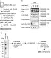
The 26S proteasome is purified together with known proteasome-associated proteins. (A) 26S proteasomes were purified from rat skeletal muscle as described in Figure 2A but in the absence of salt. Before elution, the UBL-bound proteins were incubated in 500 µL of buffer with different salt concentrations (0, 150, 300, 500 mM NaCl). The salt extract (SW) was collected, and after reequilibration in salt-free buffer, the 26S proteasomes were eluted in the 500 µL buffer containing His10-UIM. (B) Equal volumes of 26S eluates were loaded onto 3.5% native gels and analyzed by LLVY-overlay assay and Western blot as indicated. The Western blot with anti-USP14 was performed on a SDS gel (*). Doubly capped, singly capped, and PA200 hybrid proteasomes were identified as indicated. No 19S nor free 20S was detected (data not shown). (C) Mass spectrometric analysis of proteins isolated with the GST-UBL in the absence of NaCl. For a complete list of all mass spectrometric data, see Supporting Information Table 2.
Figure 4
Figure 4
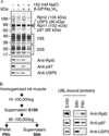
In the absence of salt, USP5 and p97 complexes are purified in large amounts together with the 26S and bind to the UBL domain directly. (A) UBL-affinity preparations were carried out in the presence or absence of 150 mM NaCl and 25 mM β-glycerophosphate (β-GP)/1 mM Na3VO4. Samples containing equal amounts of proteasomal peptidase activity were analyzed by SDS–PAGE (upper panel) and Western blot (lower panels). The single bands corresponding to Rpn1, Rpn2, USP5, and p97 were identified by mass spectrometry. (B) A crude cell extract derived from 2 g of rat skeletal muscle was centrifuged for 1 h at 100000g. The pellet was discarded and the supernatant (S100) ultracentrifuged for another 6 h at 100000g. S100, S6h, and P6h were each subjected to UBL-affinity purifications and analyzed by Western blot with the indicated antibodies.
Figure 5
Figure 5
Ubiquitin conjugates copurify with 26S and p97 complexes and are partially removed with the His10-UIM in the last purification step. (A) Proteins were isolated with the UBL domain from rat muscle as illustrated in Figure 3A. The same amount of UBL and salt wash (SW) fractions were separated by SDS–PAGE and analyzed by Western blot with anti-ubiquitin conjugate antibody. (B) To retrieve ubiquitin conjugates, His10-UIM was eluted from the Ni-NTA in binding buffer supplemented with 500 mM imidazole as illustrated by the scheme. One microgram of UBL-bound proteins from three different purifications and 1/10th of the corresponding UIM eluate were separated on SDS–PAGE and analyzed by Western blot with an anti-ubiquitin conjugate antibody. (C) UBL purifications from rat muscle were carried out in the presence or absence of 150 mM NaCl. Equal amounts of UBL- and UIM-bound proteins were analyzed by Western blot as indicated.
Figure 6
Figure 6
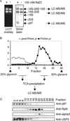
Identification of proteins associated with the 26S proteasome. (A) UBL purifications from rat muscle were carried out in the presence or absence of 150 mM NaCl. Samples containing equal amounts of proteasomal peptidase activity were separated on native gels. Proteasomes were detected by Suc-LLVY-amc overlay assay (left panel). The right panel presents a silver stain of the respective gel. The silver-stained bands of the indicated complexes were analyzed by LC-MS/MS. (B) About 100 µg of protein after UBL purification were separated on a 25–50% glycerol gradient by ultracentrifugation (22 h, 100000g). Individual fractions were analyzed for proteasomal peptidase activity (Suc-LLVY-amc) and ATPase activity (Malachite green assay). Proteins in the fractions were TCA-precipitated and subjected to mass spectrometric analysis or (C) Western blot. For a complete list of all mass spectrometric data, see Supporting Information Table 2.

Similar articles

Cited by

References

    1. Ciechanover A. Proteolysis: from the lysosome to ubiquitin and the proteasome. Nat. Rev. Mol. Cell Biol. 2005;6:79–87. - PubMed
    1. Smith DM, Benaroudj N, Goldberg A. Proteasomes and their associated ATPases: a destructive combination. J. Struct. Biol. 2006;156:72–83. - PubMed
    1. Schmidt M, Hanna J, Elsasser S, Finley D. Proteasome-associated proteins: regulation of a proteolytic machine. Biol. Chem. 2005;386:725–737. - PubMed
    1. Hanna J, Finley D. A proteasome for all occasions. FEBS Lett. 2007;581:2854–2861. - PMC - PubMed
    1. Glickman MH, Raveh D. Proteasome plasticity. FEBS Lett. 2005;579:3214–3223. - PubMed

Publication types

MeSH terms