. 2009 Mar;28(3):103-8.
doi: 10.1089/dna.2008.0792.

Generation of recombinant vaccinia viruses via green fluorescent protein selection

Affiliations

Generation of recombinant vaccinia viruses via green fluorescent protein selection

Sergei Popov et al. DNA Cell Biol. 2009 Mar.

Abstract

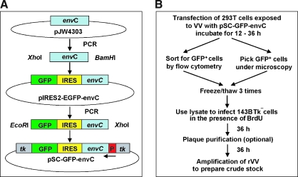
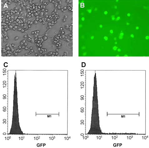
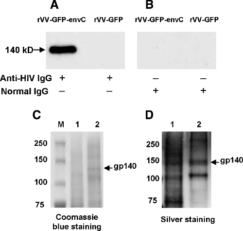
We developed a rapid method to generate recombinant vaccinia viruses (rVVs) based upon a bicistronic cassette encoding the gene for green fluorescent protein (GFP) and a foreign gene of interest separated by an internal ribosome entry site (IRES). As proof-of-concept, we inserted a mutant env gene of human immunodeficiency virus (HIV) into the cassette, which was cloned into the vaccinia virus (VV) insertion vector pSC59 under the control of the early-late VV synthetic promoter and flanked by disrupted tk gene sequences. To generate rVVs, 293T cells were inoculated with wild-type (wt) VV, followed by transfection of the modified pSC59 vector containing the bicistronic cassette, which allows expression of GFP and the protein of interest. Next, GFP-positive cells were isolated by flow cytometry or by picking under a fluorescent microscope. Thymidine kinase-deficient (Tk(-)) 143B cells were then exposed to lysates of GFP-positive 293T cells and cultured in the presence of bromodeoxyuridine. This selection allows only Tk(-) rVV to remain viable. We demonstrated the success of this GFP selection strategy by expressing high levels of mutant HIV Env. Our approach shortens the time needed to generate rVVs and represents a practical approach to generate recombinant proteins.


Figures

FIG. 1.
FIG. 1.
Strategy to generate insertion vector for rapid selection of rVVs. (A) The sequence of the mutated HIV clade C env gene, termed envC, was obtained by PCR (blue box) and cloned into the XhoI and BamHI sites of the vector pIRES2-GFP. In the second step, the sequence of the expression cassette envC–IRES–GFP was PCR amplified and cloned into vector pCS59 (sites XhoI–EcoRI) under the control of strong synthetic early/late VV promoter (red box). Gray boxes, flanking sequences of the tk gene used for targeting the insertion vector and thereby disrupt the normal VV tk gene. As a consequence, rVVs containing the desired insert are no longer able to express thymidine kinase, and thus cells infected with rVVs become resistant to the toxic effects of BrdU. (B) Experimental protocol to accelerate the generation and purification of rVVs. First, 293T cells are exposed to wt VV, followed by transfection of the insertion vector encoding the gene of interest (envC in our example). After 12 h, the GFP-positive cells are selected using flow cytometry. As an alternative to FACS sorting, rVVs can also be isolated by picking of green 293T cell plaques under a fluorescent microscope. After freezing and thawing, the GFP-positive 293T cell lysates are used to infect 143B Tk cells in the presence of BrdU (the latter prevents replication of wt VV). To obtain pure clonal rVV stocks, several rounds of plaque purification can be performed. Finally, the amplified rVVs are collected and frozen in aliquots.
FIG. 2.
FIG. 2.
Selection of rVVs using flow cytometry. (A) Phase-contrast microscopy and (B) fluorescent microscopy of 293T cells infected with rVVs expressing HIV-1 clade C envelope glycoproteins (EnvC) and GFP. Flow cytometry histograms of 293T cells infected with (C) wt VV (negative control) and (D) rVV–GFP–envC. The brightest cells were sorted and cloned by limiting dilution; viral particles were released by repeated cycles of freezing–thawing and used for infection of 143B Tk in the presence of BrdU.
FIG. 3.
FIG. 3.
Expression and purification of EnvC from 143B cells infected with rVV–GFP–envC. (A–B) Infected cells were solubilized in Laemmli buffer and fractionated in gradient PAGE gel (4–12%); proteins were transferred to the PVDF membrane and probed with (A) anti-HIV-1 human IgG isolated from serum of an HIV-1–infected patient and (B) normal human IgG. (C–D) Analysis of purified mutant HIV envelope glycoproteins produced in 143B cells infected with rVV–GFP–envC. The mutant HIV EnvC yielded the expected band of approximately 140 kDa under denaturing conditions. (C) SDS-PAGE Coomassie blue staining. Lane 1, 10 μg of total protein from mock-infected cells; lane 2, 10 μg of protein purified by lentil lectin affinity column and Superdex-S200 size exclusion column chromatography using extracts of cells infected with rVV–GFP–envC. (D) Silver staining. Lane 1, total protein (2 μg) after the lentil lectin affinity column purification; lane 2, purified protein (2 μg) after size exclusion column chromatography purification.

Comment in

References

    1. Bleckwenn N.A. Bentley W.E. Shiloach J. Exploring vaccinia virus as a tool for large-scale recombinant protein expression. Biotechnol Prog. 2003;19:130–136. - PubMed
    1. Borman A.M. Le Mercier P. Girard M. Kean K.M. Comparison of picornaviral IRES-driven internal initiation of translation in cultured cells of different origins. Nucleic Acids Res. 1997;25:925–932. - PMC - PubMed
    1. Chakrabarti S. Sisler J.R. Moss B. Compact, synthetic, vaccinia virus early/late promoter for protein expression. Biotechniques. 1997;23:1094–1097. - PubMed
    1. Davison A.J. Moss B. Structure of vaccinia virus early promoters. J Mol Biol. 1989a;210:749–769. - PubMed
    1. Davison A.J. Moss B. Structure of vaccinia virus late promoters. J Mol Biol. 1989b;210:771–784. - PubMed

Publication types