. 2009 Apr 21;48(15):3354-69.
doi: 10.1021/bi802029t.

Role of conserved tyrosine residues in NiSOD catalysis: a case of convergent evolution

Affiliations

Role of conserved tyrosine residues in NiSOD catalysis: a case of convergent evolution

Robert W Herbst et al. Biochemistry. .

Abstract

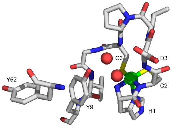
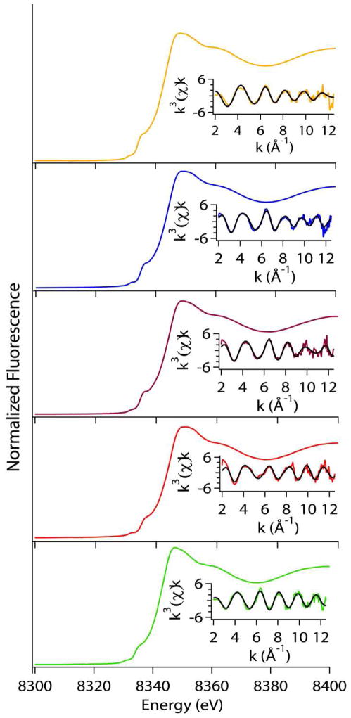
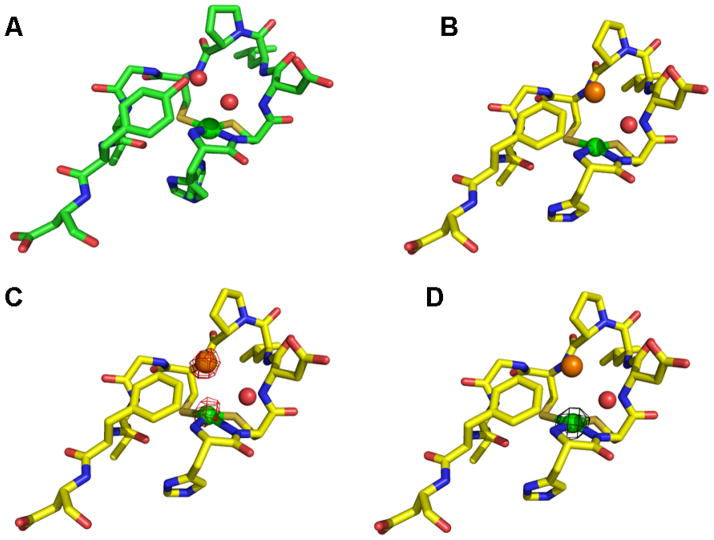
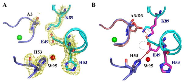
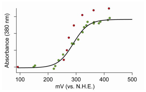
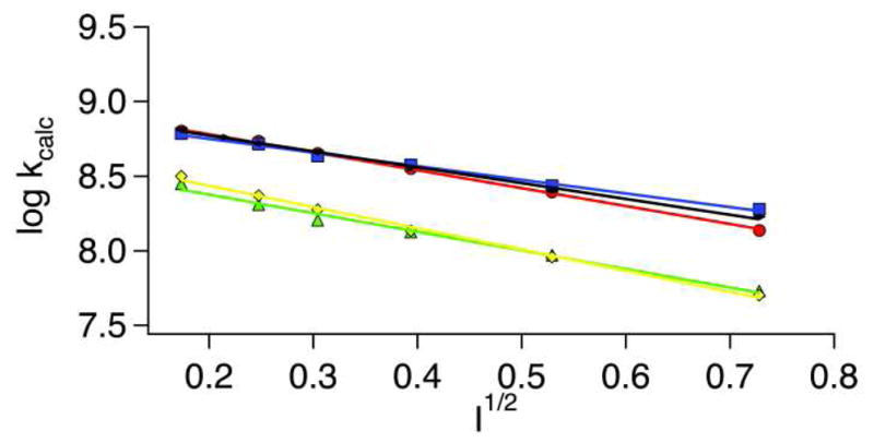
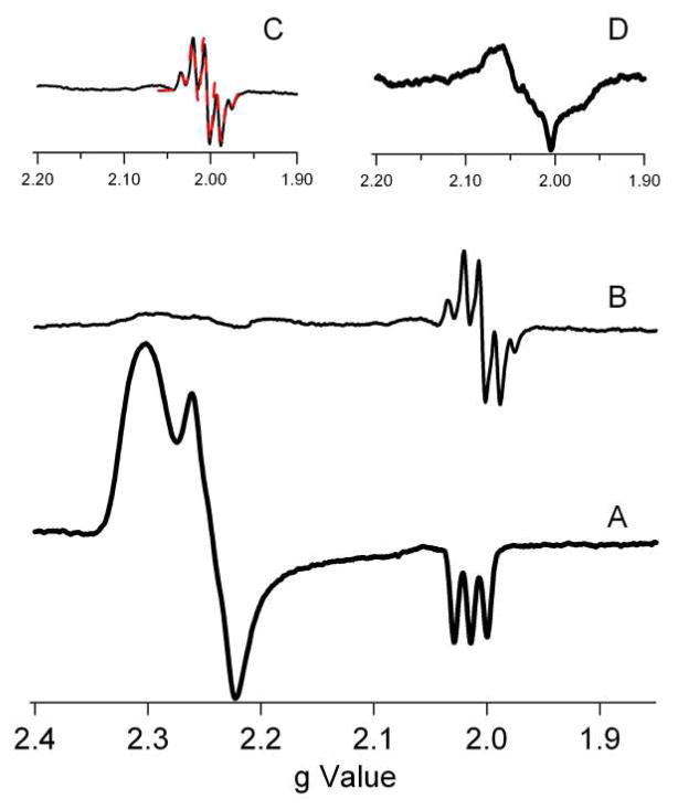
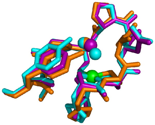
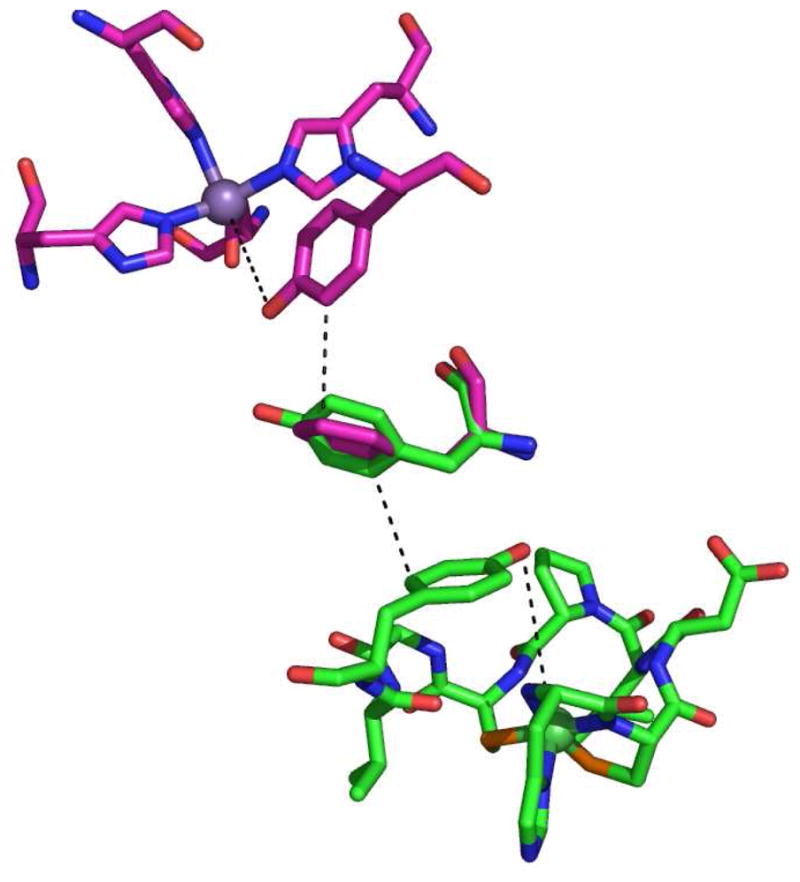
Superoxide dismutases rely on protein structural elements to adjust the redox potential of the metallocenter to an optimum value near 300 mV (vs NHE), to provide a source of protons for catalysis, and to control the access of anions to the active site. These aspects of the catalytic mechanism are examined herein for recombinant preparations of the nickel-dependent SOD (NiSOD) from Streptomyces coelicolor and for a series of mutants that affect a key tyrosine residue, Tyr9 (Y9F-, Y62F-, Y9F/Y62F-, and D3A-NiSOD). Structural aspects of the nickel sites are examined by a combination of EPR and X-ray absorption spectroscopies, and by single-crystal X-ray diffraction at approximately 1.9 A resolution in the case of Y9F- and D3A-NiSODs. The functional effects of the mutations are examined by kinetic studies employing pulse radiolytic generation of O2- and by redox titrations. These studies reveal that although the structure of the nickel center in NiSOD is unique, the ligand environment is designed to optimize the redox potential at 290 mV and results in the oxidation of 50% of the nickel centers in the oxidized hexamer. Kinetic investigations show that all of the mutant proteins have considerable activity. In the case of Y9F-NiSOD, the enzyme exhibits saturation behavior that is not observed in wild-type (WT) NiSOD and suggests that release of peroxide is inhibited. The crystal structure of Y9F-NiSOD reveals an anion binding site that is occupied by either Cl- or Br- and is located close to but not within bonding distance of the nickel center. The structure of D3A-NiSOD reveals that in addition to affecting the interaction between subunits, this mutation repositions Tyr9 and leads to altered chemistry with peroxide. Comparisons with Mn(SOD) and Fe(SOD) reveal that although different strategies for adjusting the redox potential and supply of protons are employed, NiSOD has evolved a similar strategy for controlling the access of anions to the active site.


Figures

Figure 1
Figure 1
Active site from one monomer of the homohexameric NiSOD from Streptomyces coelicolor, showing the first nine residues and Tyr62(17). The nickel is shown in the five-coordinate pyramidal oxidized His-on form. PDB code 1T6U. Image was generated in PyMOL (75).
Figure 2
Figure 2
Comparison of XAS data for Tyrosine mutant NiSODs; WT(Yellow), Y9F(Blue), Y62F(Purple), Y9FY62F(Red), D3A(Green). Left: Normalized XANES spectra. Right: Unfiltered EXAFS data in solid colored line and fits (black) from Table 6. The range of the k-space fits is 2–12.5 Å−1 for all data.
Figure 3
Figure 3
NiSOD hook domain. A. WT-NiSOD N-terminal region showing the active site Ni ion (green sphere) and two water molecules (red spheres). B. The same region of the Y9F-NiSOD mutant. A chloride ion (orange sphere) at the anion-binding site replaces a water molecule. C. The same region of the Y9F-NiSOD(Br) structure. An anomalous difference map (red cage), contoured at 4σ, from data measured above the bromide absorption edge energy. Only the bromide and nickel show an anomalous signal. D. The same region of the Y9F-NiSOD(Cl) structure. An anomalous difference map (black cage), contoured at 4σ, from data measured above the bromide absorption edge. Only the nickel shows an anomalous signal. The Ni centers are modeled as the “His-off” state, the actual state is most likely a mixture of “His-on” and “His-off”, but higher B-factors led to poorly resolved electron density at the N-terminus. Images were generated in PyMOL (75).
Figure 4
Figure 4
Salt bridge changes in the D3A-NiSOD mutant. A. The D3A-NiSOD mutant is shown at the interface between neighboring monomers (in blue and cyan) in the hexamer. A weighted 2Fo-Fc map shows the electron density in the interface. B. Superposition of this interface from D3A-NiSOD (in blue and cyan) onto wild type (pink and magenta) shows the movement of Glu49 and Lys89. Images were generated in PyMOL (75).
Figure 5
Figure 5
Redox titrations of WT-NiSOD. The green points are the initial reduction of oxidized to reduced NiSOD and the red points were obtained by re-oxidation of the same sample. The black line depicts the best fit of the reductive titration (green dots) using equation 4.
Figure 6
Figure 6
Ionic strength dependence of NiSOD catalysis. Y9F- and WT-NiSOD were reacted with both NaCl (red-WT and yellow-Y9F) and NaClO4 (blue-WT and green-Y9F), while Y62F was only reacted with NaCl (black). Pulse radiolytic conditions were the same as those mentioned in Materials and Methods, except 0.5 M – 0.0156 M of either NaCl or NaClO4 was added to the solution being monitored to probe the effect of ionic strength.
Figure 7
Figure 7
X-band EPR data from D3A-NiSOD. (A) As-isolated D3A. (B) H2O2 treated D3A. (C) Enlargement of the region near g = 2.00 expanded (solid line) and a simulation with parameters gx=gy=gz=2.004, Axx=Ayy=5 G, Azz=20 G (dashed line-red). (D) H2O2 treated D3A-NiSOD (d4-Tyr), showing alteration of hyperfine due to tyrosine ring protons. All spectra are shown with the same intensity scale with the exception of D, which has a scale half as large.
Figure 8
Figure 8
Superposition of the Ni-hook domains of WT- (blue), Y9F- (purple) and D3A- (Orange) NiSODs showing the position of Y9 in each. Image was generated in PyMOL (75).
Figure 9
Figure 9
Superposition of the active site structures of hMnSOD (pink) and S. coelicolor NiSOD (green) showing the position of aromatic residues. Image was generated in PyMOL (75).
Scheme 1
Scheme 1
Proposed outer-sphere mechanism for NiSOD. Ni(III) is indicated by the His-on complex, while Ni(II) is denoted as a His-off complex, although removal of the His1 during turnover may not occur. Superoxide is denoted by the orange molecule and water is denoted by red spheres. Images were generated in PyMOL (75).

References

    1. Cabelli DE, Riley D, Rodriguez JA, Valentine JS, Zhu H. Models of Superoxide Dismutases. In: Meunier B, editor. Biomimetic Oxidations Catalyzed by Transition Metal Complexes. Ch 10 1998.
    1. Fridovich I. Superoxide radical and superoxide dismutases. Annu Rev Biochem. 1995;64:97–112. - PubMed
    1. Miller AF, Sorkin D. Superoxide Dismutases: A Molecular Perspective. Comments Mol Cell Biophys. 1997;9:1–48.
    1. Touati D. Superoxide Dismutases in Bacteria and Pathogen Protists. In: Scandalios JG, editor. Oxidative Stress and the Molecular Biology of Antioxidant Defenses. Cold Spring Harbor Laboratory Press; NY: 1997. pp. 447–493.
    1. Lee JW, Roe JH, Kang SO. Nickel-containing superoxide dismutase. Superoxide Dismutase. 2002:90–101. - PubMed

Publication types

MeSH terms