. 2009 Mar;108(6):1550-60.
doi: 10.1111/j.1471-4159.2009.05901.x. Epub 2009 Jan 22.

Chronic copper exposure exacerbates both amyloid and tau pathology and selectively dysregulates cdk5 in a mouse model of AD

Affiliations

Chronic copper exposure exacerbates both amyloid and tau pathology and selectively dysregulates cdk5 in a mouse model of AD

Masashi Kitazawa et al. J Neurochem. 2009 Mar.

Abstract

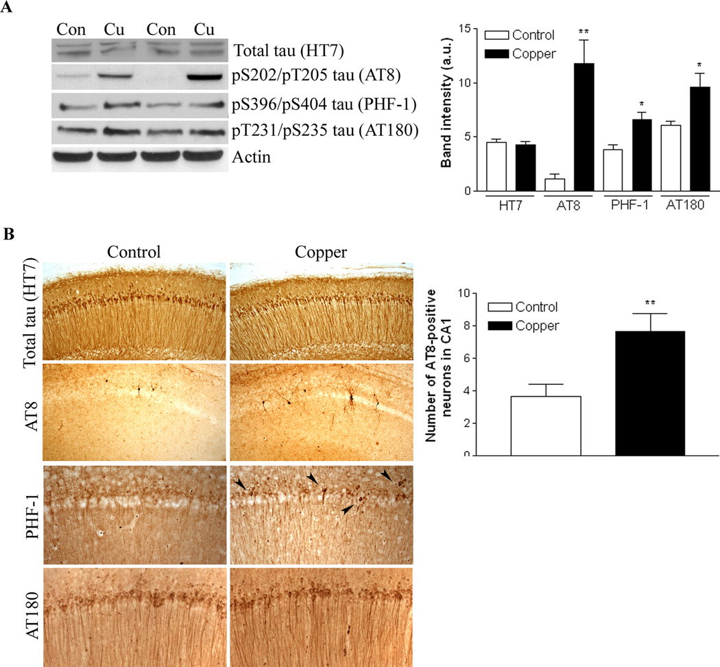
Excess copper exposure is thought to be linked to the development of Alzheimer's disease (AD) neuropathology. However, the mechanism by which copper affects the CNS remains unclear. To investigate the effect of chronic copper exposure on both beta-amyloid and tau pathologies, we treated young triple transgenic (3xTg-AD) mice with 250 ppm copper-containing water for a period of 3 or 9 months. Copper exposure resulted in altered amyloid precursor protein processing; increased accumulation of the amyloid precursor protein and its proteolytic product, C99 fragment, along with increased generation of amyloid-beta peptides and oligomers. These changes were found to be mediated via up-regulation of BACE1 as significant increases in BACE1 levels and deposits were detected around plaques in mice following copper exposure. Furthermore, tau pathology within hippocampal neurons was exacerbated in copper-exposed 3xTg-AD group. Increased tau phosphorylation was closely correlated with aberrant cdk5/p25 activation, suggesting a role for this kinase in the development of copper-induced tau pathology. Taken together, our data suggest that chronic copper exposure accelerates not only amyloid pathology but also tau pathology in a mouse model of AD.


Figures

Figure 1
Figure 1. 3-month copper exposure alters APP processing
(A) Immunoblot analysis of APP, C99, C83, BACE1, ADAM10 (full-length, ~100 kDa) and PS1 in brain homogenates from control or copper-exposed mice (n=10 per group). β-actin levels are used as a loading control. Densitometric analyses of steady-state levels of (B) APP, (C) C99 and C83, and (D) BACE1 and ADAM10 are presented in graph with mean ± S.E.M. (*p<0.05 compared to control group). (E) Total Aβ levels from detergentsoluble and –insoluble fractions are measured by ELISA (n=10 per groups). *p<0.05 compared to control group. (F) Representative immunohistochemical staining of APP/Aβ by 6E10 antibody.
Figure 2
Figure 2. 3-month copper exposure does not exhibit pathological tau accumulation
(A) Immunoblot analysis of total tau (HT7) and various phospho-tau epitopes including pS202/pT205 tau (AT8), pS396/pS404 tau (PHF-1) and pT231/pS235 tau (AT180) in brain homogenates from control or copper-exposed mice (n=10 per group). β-actin levels are used as a loading control. Densitometric analyses are shown in the graph (mean ± S.E.M). No significant differences of total tau or phospho-tau levels are detected between control and copper-exposed mice. (B) Representative immunohistochemical staining of tau in hippocampus region. AT8-positive neurons in CA1 hippocampus are counted and and determined the significance in the graph shown on the right panel (n=10 per group, *p<0.05 compared to control).
Figure 3
Figure 3. 9-month copper exposure exacerbates Aβ pathology in the brain
(A) Immunoblot analysis of APP, C99, C83, BACE1, ADAM10 (full-length, ~100 kDa) and PS1 in brain homogenates from control or copper-exposed group (n=10 per group). β-actin levels are used as a loading control. Densitometric analyses of steady-state levels of (B) APP, (C) C99 and C83, and (D) BACE1 and ADAM10 are presented in graph (*p<0.05 or **p<0.01 compared to control group). Total Aβ levels from (E) detergent-soluble and (F) detergent-insoluble fractions are measured by ELISA (n=10 per groups). *p<0.05 compared to control group. (G) Representative immunohistochemical staining of APP/Aβ by 6E10, Aβ40 and Aβ42 specific antibodies.
Figure 4
Figure 4. Detection of oligomeric Aβ species in the brain
(A) Representative double immunofluorescent staining of thioflavin S-positive amyloid plaques and A11-positive amyloid oligomers in the brains of control and 9-month copper-exposed mice. Dot blot analysis of A11-positive oligomers demosntrates a marginal increase in copper-exposed mice (n=6 per group). (B) Representative double immunofluorescent staining of astrocytic marker, GFAP and A11-positive amyloid oligomers in the brain of 3xTg-AD mice. Arrowheads represent colocalization of GFAP and A11-positive staining, indicating A11-positive oligomers are present in the astrocytes.
Figure 5
Figure 5. 9-month copper exposure exacerbates tau pathology
(A) Immunoblot analysis of total tau (HT7) and phospho-tau epitopes including pS202/pT205 tau (AT8), pS396/pS404 tau (PHF-1) and pT231/pS235 tau (AT180) in brain homogenates from control or copper-exposed mice (n=10 per group). β-actin levels are used as a loading control. Densitometric analyses reveal significant increases in brain phospho-tau levels in copper-exposed mice (mean ± S.E.M, *p<0.05 or **p<0.01 compared to control). (B) Representative immunohistochemical staining of tau in hippocampus region. AT8-positive neurons in CA1 hippocampus are counted and determined the significance in the graph shown on the right panel (n=10 per group, **p<0.01 compared to control). The arrowheads in PHF-1 staining show highly condensed somatodendritic accumulation of phospho-tau, suggestive of late pathological tau aggregates in CA1 hippocampus of copper-exposed mice.
Figure 6
Figure 6. Copper exposure activates cdk5 via increased generation of p25
(A) Cdk5 and GSK-3β activation states were determined by immunoblotting of cdk5, p35/p25, phospho-GSK-3β (at serine 9), and total GSK-3β in 3-month and 9-month copper-exposed group and control group (n=10 per group). (B) Densitometric analysis of p25 fragment is shown in the graph (mean ± S.E.M., **p<0.01 compared to age-matched control). A significant increase in the formation of p25 is detected in 9-month copper-exposed mice. (C) Brain calpain activity of 9-month copper-exposed mice is measured using a specific fluorogenic substrate. The calpain activity is expressed as relative fluorescent unit (R.F.U.), and the graph represents mean ± S.E.M. of control (n=4) and copper-exposed mice (n=6).
Figure 7
Figure 7. Brain interaction of BACE1 or SOD1 with CCS is altered in chronic copper exposure
Steady-state levels of CCS and SOD1 were determined by immunoblotting (n=5–6 per group). The interaction of BACE1 or SOD1 with CCS was determined by immunoprecipitating CCS followed by immunoblotting with BACE1 or SOD1. Densitometric analysis showed a significance at *p<0.05 or **p<0.01 compared to agematched control group (n=5 per group).

References

    1. Angeletti B, Waldron KJ, Freeman KB, Bawagan H, Hussain I, Miller CC, Lau KF, Tennant ME, Dennison C, Robinson NJ, Dingwall C. BACE1 cytoplasmic domain interacts with the copper chaperone for superoxide dismutase-1 and binds copper. J Biol Chem. 2005;280:17930–17937. - PubMed
    1. Armendariz AD, Gonzalez M, Loguinov AV, Vulpe CD. Gene expression profiling in chronic copper overload reveals upregulation of Prnp and App. Physiol Genomics. 2004;20:45–54. - PubMed
    1. Atwood CS, Scarpa RC, Huang X, Moir RD, Jones WD, Fairlie DP, Tanzi RE, Bush AI. Characterization of copper interactions with alzheimer amyloid beta peptides: identification of an attomolar-affinity copper binding site on amyloid beta1-42. J Neurochem. 2000;75:1219–1233. - PubMed
    1. Atwood CS, Moir RD, Huang X, Scarpa RC, Bacarra NM, Romano DM, Hartshorn MA, Tanzi RE, Bush AI. Dramatic aggregation of Alzheimer abeta by Cu(II) is induced by conditions representing physiological acidosis. J Biol Chem. 1998;273:12817–12826. - PubMed
    1. Augustinack JC, Sanders JL, Tsai LH, Hyman BT. Colocalization and fluorescence resonance energy transfer between cdk5 and AT8 suggests a close association in pre-neurofibrillary tangles and neurofibrillary tangles. J Neuropathol Exp Neurol. 2002;61:557–564. - PubMed

Publication types

MeSH terms