. 2009 Feb;13(2):295-308.
doi: 10.1111/j.1582-4934.2008.00668.x. Epub 2009 Jan 27.

A distinct type of cell in myocardium: interstitial Cajal-like cells (ICLCs)

Affiliations

A distinct type of cell in myocardium: interstitial Cajal-like cells (ICLCs)

S Kostin et al. J Cell Mol Med. 2009 Feb.

Abstract

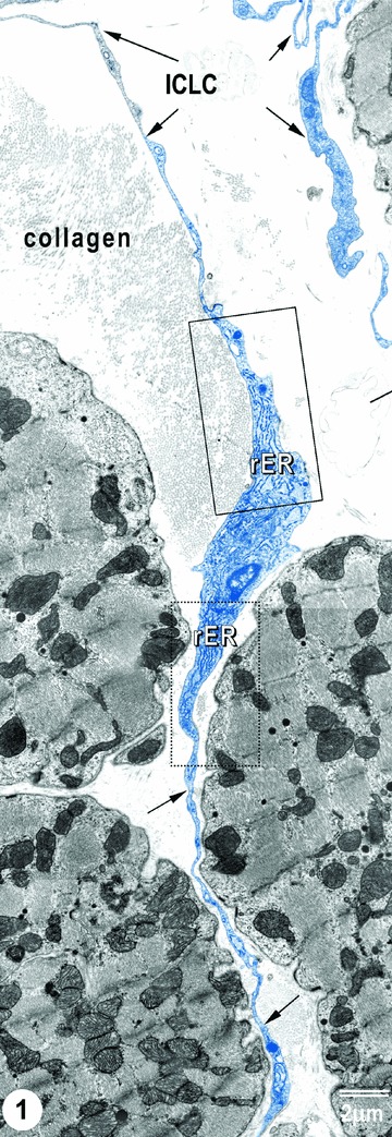
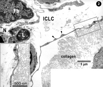
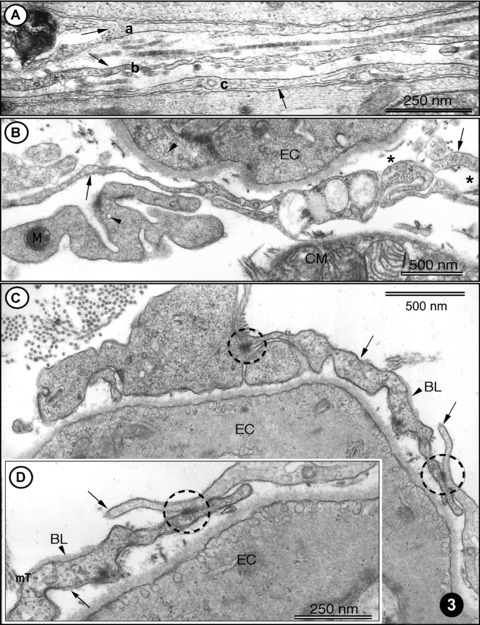
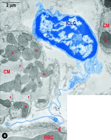
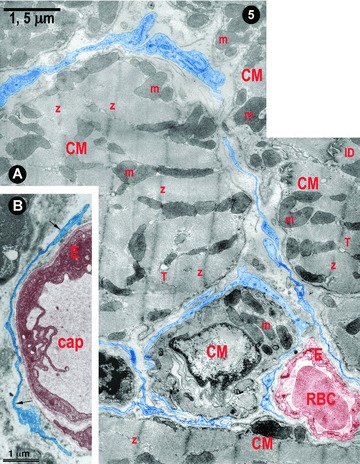
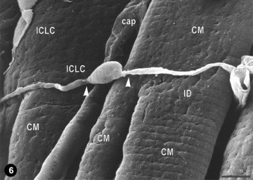
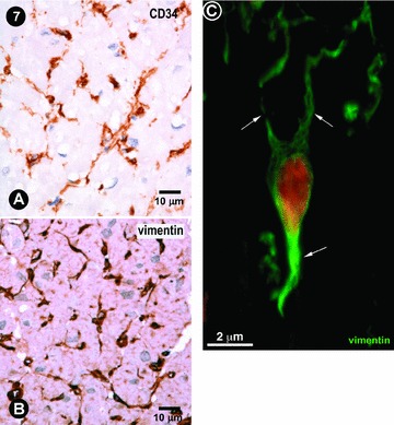
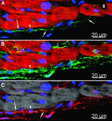
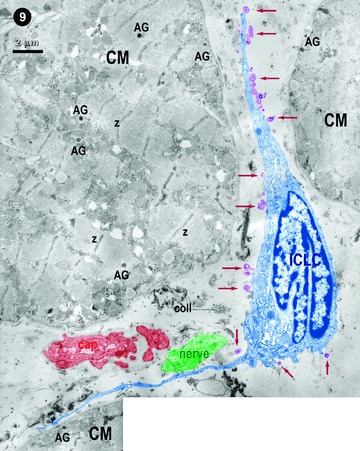
The existence of a novel type of interstitial cells in the heart, interstitial Cajal-like cells (ICLCs), had been described for the first time in 2005. Their identification was mainly based on ultrastructural criteria: very long (tens up to hundreds of micrometres) and moniliform prolongations, which are extremely thin (less than 0.2 microm), below the resolving power of light microscopy. Myocardial ICLCs were also identified by methylene-blue vital staining, silver impregnation, and immunoreactivity for CD 34, vimentin, CD117/c-kit, etc. Although a series of studies provided evidence for the existence of ICLCs in human atria and rat ventricles, further investigations in other laboratories, using additional techniques, are required to substantiate the consistency of these findings. Here we provide further evidence for the existence of ICLCs in human and mammalian hearts (by transmission and scanning electron microscopy, as well as confocal laser scanning microscopy). Noteworthy, we confirm that ICLCs communicate with neighbouring cells via shedding (micro)vesicles. Although these so-called ICLCs represent a distinct type of cells, different from classical interstitial cells of Cajal, or fibroblasts, their role(s) in myocardium remain(s) to be established. Several hypotheses are proposed: (i) adult stromal (mesenchymal) stem cells, which might participate in cardiac repair/remodelling; (ii) intercellular signalling (e.g. via shedding microvesicles); (iii) chemo-mechanical transducers and (iv) players in pacemaking and/or arrhytmogenesis, and so on.


Figures

1
1
TEM image of ICLCs (rat) in the right atrial interstitium. ICLCs are indicated by arrows. Note the characteristic aspects of ICLC processes: very long and very thin cellular elongations, with uneven calibre (moniliform aspect). Insets show portions of ICLC cell body, containing (abundant) rough endoplasmic reticulum.
2
2
Higher magnification of the upper part of Fig. 1. ICLC processes appear very thin with dilations (arrowheads). ICLC extensions are located in close vicinity with lymphocytes (L), nerves (N) and (presumably) fibroblasts (Fb). The inset shows a higher magnification image.
3
3
(A)-(D) TEM images of rat left ventricular myocardium. (A) Three ICLC processes (arrows: a, b, c) are located in the myocardial interstitium. Note the cross-striated pattern of collagen fibrils (in the middle part). (B) A typical example of ICLC processes (arrows) located in vicinity of endothelial cells (EC) and cardiomyocytes. The presence of caveolae (arrowheads) is a typical feature of ICLCs and ECs. Note the cross-section of intermediate filaments (asterisk) inside ICLC processes. (C) ICLCs (arrows) located in close vicinity of endothelial cells (EC). Cell-to-cell contacts of ICLCs are indicated with circles. Note the presence of the basal lamina (BL, arrowhead), which can occasionally be observed in ICLCs. (D) Higher magnification of the right part of (C) showing jonctional complexes (circle) between two ICLCs, indicated with arrows. BL – basal lamina. Cross-sectioned micro-tubules (mT) can be seen in ICLC processes.
4
4
Digitally coloured electron micrograph of rat ventricular myocardium: a typical ICLCs (blue) with a long tortuous process and with uneven calibre (moniliform) in between two cardiomyocytes. ICLC prolongation is in the vicinity of a blood capillary; m – mitochondria, Z–Z band, RBC – red blood cell, E – endothelium. The circle indicates a dilation of the ICLC process, which contains the so-called ‘Ca2+ Handling Unit’ composed of: one to two mitochondria, one to two elements of ER and one or a few caveolae.
5
5
(A) and (B) Digitally coloured electron micrographs of rat (A) and human (B) ventricular myocardium: typical ICLC processes (blue) forming a network in the narrow spaces between cardiomyocytes (A) and surrounding blood capillaries (A and B). m – mitochondria, Z–Z band, ID – intercalated disc, T – T tubules (cross-section), RBC – red blood cell, E – endothelium and cap – capillary.
6
6
Representative scanning electron micrograph (monkey left ventricular myocardium) showing a typical ICLC located across the cardiomyocytes. Another (possible) ICLC appears located along the cardiomyocytes (upper left). The three-dimensional vision reveals close interconnections of ICLCs with cardiomyocytes and capillaries (cap); compare with Fig. 5. Note that ICLC processes begin from the cell body abruptly, as very thin prolongations (arrow heads); ID.
7
7
(A) and (B) Usual immunocytochemistry of human left atrial myocardium. Some cells, with characteristic morphologic features for ICLCs, appear immunopositive for CD34 and vimentin. Mayer's haematolxylin counter-stain for nuclei. (C) Human left ventricular myocardium: CLSM of a vimentin positive ICLCs (green) with at least three processes (arrows). Nucleus is stained red with 7-aminoactinomycin D.
8
8
Immunohistochemistry of human left ventricle myocardium; CLSM. (A) CD117/c-kit+ cells (green) with long cytoplasmic processes (indicated by arrows) indicated by arrows; myofibrils are stained bright-red with Alexa 633-phalloidin and nuclei are stained blue with DAPI. The arrowhead shows the nucleus of a c-kit+ cell. (B) Vimentin positive cells (green); myofibrils appear bright red, stained with Alexa 633-phalloidin and nuclei blue with DAPI (arrowhead). (C) Merged view of images (A) and (B) showing that c-kit signal (green) co-localizes with vimentin (red) resulting a yellow colour (arrows). Note that most of cells are only vimentin positive (red) and only a few are positive for both c-kit and vimentin. Myofibrils are pseudo-coloured in grey. The arrowhead indicates a nucleus (blue).
9
9
Digitally coloured transmission electron micrograph: oblique section of human right atrial myocardium. An ICLC (blue) in between cardiomyocytes, but in proximity of a blood capillary (brownish) and a bundle of nerve fibres (green). The ICLC is shedding (micro)vesicles (violet) budding from the body of the ICLC and its processes (arrows). Z–Z bands; AG – atrial granules.

References

    1. Rumessen JJ, Vanderwinden JM. Interstitial cells in the musculature of the gastrointestinal tract: Cajal and beyond. Int Rev Cytol. 2003;229:115–208. - PubMed
    1. Faussone-Pellegrini MS. Interstitial cells of Cajal: once negligible players, now blazing protagonists. Ital J Anat Embryol. 2005;110:11–31. - PubMed
    1. Sanders KM, Koh SD, Ward SM. Interstitial cells of Cajal as pacemakers in the gastrointestinal tract. Annu Rev Physiol. 2006;68:307–43. - PubMed
    1. Nakayama S, Kajioka S, Goto K, Takaki M, Liu HN. Calcium-associated mechanisms in gut pacemaker activity. J Cell Mol Med. 2007;11:958–68. - PMC - PubMed
    1. Yin J, Chen JD. Roles of interstitial cells of Cajal in regulating gastrointestinal motility: in vitro versus in vivo studies. J Cell Mol Med. 2008;12:1118–29. - PMC - PubMed