. 2009 Jan 30:3:42.
doi: 10.1186/1752-1947-3-42.

Two-dimensional power Doppler-three-dimensional ultrasound imaging of a cesarean section dehiscence with utero-peritoneal fistula: a case report

Affiliations

Two-dimensional power Doppler-three-dimensional ultrasound imaging of a cesarean section dehiscence with utero-peritoneal fistula: a case report

Pedro Royo et al. J Med Case Rep. .

Abstract

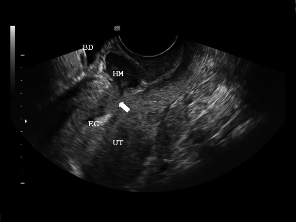
Introduction: An imaging diagnosis after an iterative cesarean delivery is reviewed demonstrating a fine ultrasound-pathologic correlation.

Case presentation: A 33-year-old woman (G3, P3) presented referring intense dysmenorrhea and intermenstrual spotting since her third cesarean delivery, 1 year before. A cesarean section dehiscence with utero-peritoneal fistula was diagnosed by transvaginal ultrasound.

Conclusion: We can conclude that transvaginal two-dimensional power Doppler and three-dimensional ultrasound are highly accurate in detecting cesarean section dehiscence and uterine fistula.


Figures

Figure 1
Figure 1
Three-dimensional transvaginal ultrasound scan (in multiplane acquisition mode) of the uterus-hematoma-bladder complex (UT, HM, BD respectively). Please note that the white pixel (placed in the center of each image) always correspond with the same space point of the three orthogonal planes, and is located referring HM, between UT (at the level of the uterine scar) and just beneath BD. Defect's surface three-dimensional reconstruction (of the coronal plane) correspond with bottom right picture, and is framed with a white arrow (instead of white pixel).
Figure 2
Figure 2
Two-dimensional transvaginal uterine (UT) ultrasound on longitudinal plane showing the communication (arrow) of the hematoma (HM) with the endometrial cavity (EC).
Figure 3
Figure 3
Intra-operative picture showing the defect on the lower uterine segment after dissection (arrow).
Figure 4
Figure 4
Pathological image of the uterus showing the defect.

References

    1. Guimarães Filho HA, da Costa LL, Araujo Júnior E, Zanforlin Filho SM, Pires CR, Nardozza LM, Mattar R. Diagnosis of uteroperitoneal fistula through color Doppler hysterosonography. Arch Gynecol Obstet. 2007;276:85–86. doi: 10.1007/s00404-006-0306-0. - DOI - PubMed
    1. Porcaro AB, Zicari M, Zecchini Antoniolli S, Pianon R, Monaco C, Migliorini F, Longo M, Comunale L. Vesicouterine fistulas following cesarean section: report on a case, review and update of the literature. Int Urol Nephrol. 2002;34:335–344. doi: 10.1023/A:1024443822378. - DOI - PubMed
    1. Youssef AF. Menouria following lower segment Caesarean section. A syndrome. Am J Obstet Gynecol. 1957. pp. 759–767. - PubMed
    1. Jozwik M, Jozwik M. Clinical classification of vesicouterine fistula. Int J Gynaecol Obstet. 2000;70:353–357. doi: 10.1016/S0020-7292(00)00247-2. - DOI - PubMed
    1. Bromley B, Pitcher BL, Klapholz H, Lichter E, Benacerraf BR. Sonographic appearance of uterine scar dehiscence. Int J Gynaecol Obstet. 1995;51:53–56. doi: 10.1016/0020-7292(95)80009-2. - DOI - PubMed

LinkOut - more resources