. 2009 Jan 31:4:6.
doi: 10.1186/1749-7922-4-6.

Percutaneous retrieval of a biliary stent after migration and ileal perforation

Affiliations

Percutaneous retrieval of a biliary stent after migration and ileal perforation

Derek M Culnan et al. World J Emerg Surg. .

Abstract

We present a case of a migrated biliary stent that resulted in a distal small bowel perforation, abscess formation and high grade partial small bowel obstruction in a medically stable patient without signs of sepsis or diffuse peritonitis. We performed a percutaneous drainage of the abscess followed by percutaneous retrieval of the stent. The entero-peritoneal fistula closed spontaneously with a drain in place. We conclude, migrated biliary stents associated with perforation distal to the Ligament of Trietz (LOT), may be treated by percutaneous drainage of the abscess and retrieval of the stent from the peritoneal cavity, even when associated with a large intra-abdominal abscess.


Figures

Figure 1
Figure 1
CT Scan with right lower quadrant abscess. Computer tomography images with intravenous and oral contrast demonstrating left lower quadrant abscess and small bowel obstruction. Grey arrows denote the abscess cavity. White arrows denote the endostent.
Figure 2
Figure 2
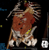
CT Scan of the common bile duct stent. 3-Dimensional reconstruction of CT data demonstrating the migrated biliary stent to be extraluminal in the left lower quadrant.
Figure 3
Figure 3
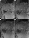
Fluroscopic images of the extraluminal biliary stent. Fluroscopic images demonstrating the retrieval of the extraluminal biliary stent. Panel A shows the catheter to be within the abscess cavity. Panel B shows the snare engaging the stent. Panel C shows the stent being removed through the sheath. Panel D shows the abscess cavity without the stent present.
Figure 4
Figure 4
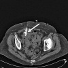
CT image of collapsed abscess cavity. CT image of the pelvis without contrast shows the drain in place and the abscess cavity completely collapsed.

References

    1. Lammer J, Neumayer K. Biliary drainage endoprostheses: experience with 201 placements. Radiology. 1986;159:625–629. - PubMed
    1. Mueller PR, Ferrucci JT, Jr, Teplick SK, vanSonnenberg E, Haskin PH, Butch RJ, Papanicolaou N. Biliary stent endoprosthesis: analysis of complications in 113 patients. Radiology. 1985;156:637–639. - PubMed
    1. Johanson JF, Schmalz MJ, Geenen JE. Incidence and risk factors for biliary and pancreatic stent migration. Gastrointest Endosc. 1992;38:341–346. doi: 10.1016/S0016-5107(92)70429-5. - DOI - PubMed
    1. Diller R, Senninger N, Kautz G, Tubergen D. Stent migration necessitating surgical intervention. Surg Endosc. 2003;17:1803–1807. doi: 10.1007/s00464-002-9163-5. - DOI - PubMed
    1. Saranga Bharathi R, Rao P, Ghosh K. Iatrogenic duodenal perforations caused by endoscopic biliary stenting and stent migration: an update. Endoscopy. 2006;38:1271–1274. doi: 10.1055/s-2006-944960. - DOI - PubMed

LinkOut - more resources