

Gut health immunomodulatory and anti-inflammatory functions of gut enzyme digested high protein micro-nutrient dietary supplement-Enprocal
- PMID: 19183498
- PMCID: PMC2667481
- DOI: 10.1186/1471-2172-10-7
Gut health immunomodulatory and anti-inflammatory functions of gut enzyme digested high protein micro-nutrient dietary supplement-Enprocal
Abstract
Background: Enprocal is a high-protein micro-nutrient rich formulated supplementary food designed to meet the nutritional needs of the frail elderly and be delivered to them in every day foods. We studied the potential of Enprocal to improve gut and immune health using simple and robust bioassays for gut cell proliferation, intestinal integrity/permeability, immunomodulatory, anti-inflammatory and anti-oxidative activities. Effects of Enprocal were compared with whey protein concentrate 80 (WPC), heat treated skim milk powder, and other commercially available milk derived products.
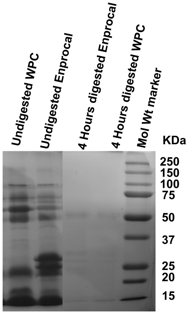
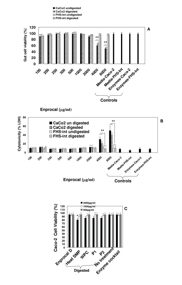
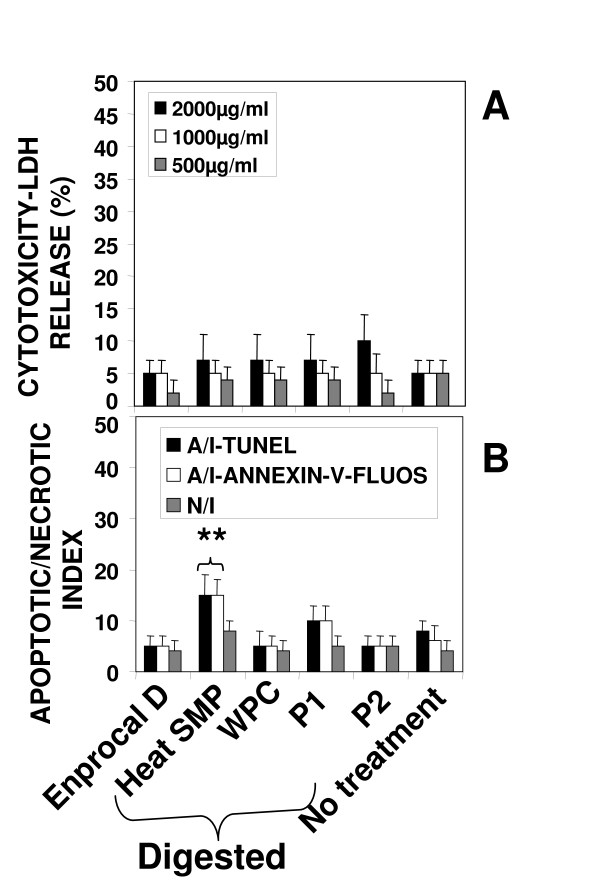
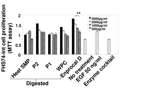
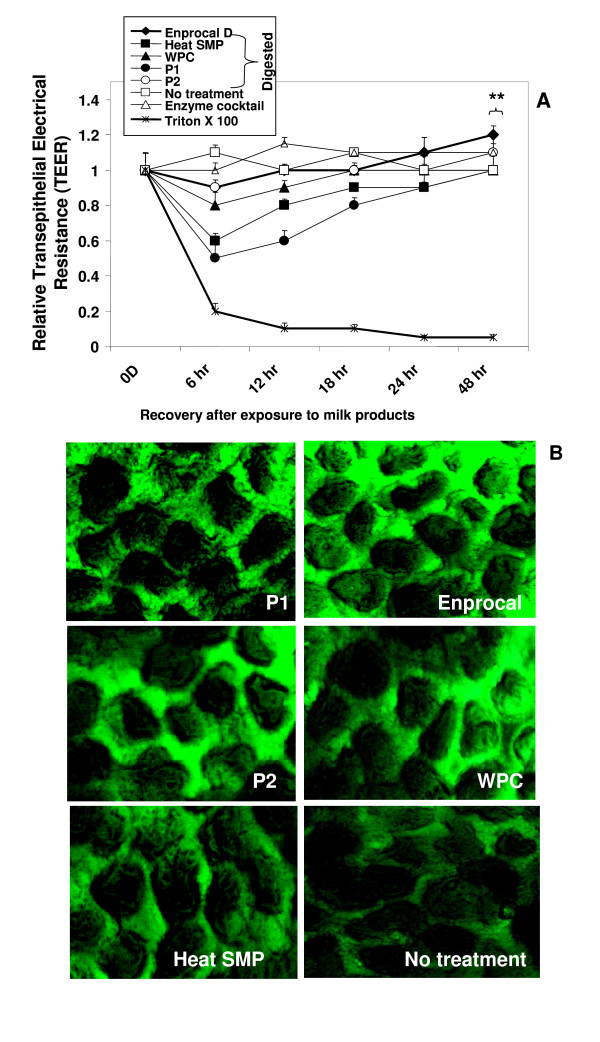
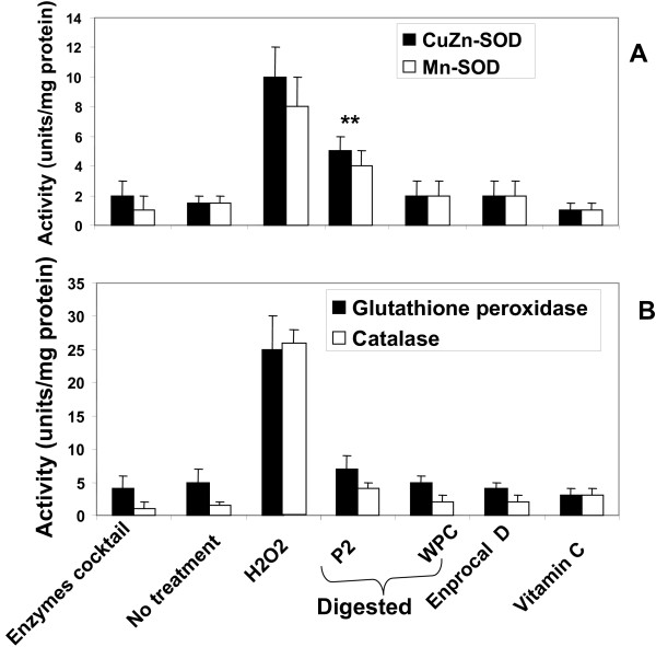
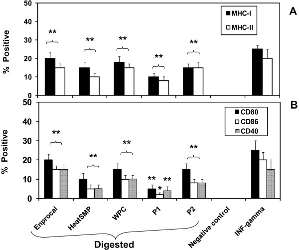
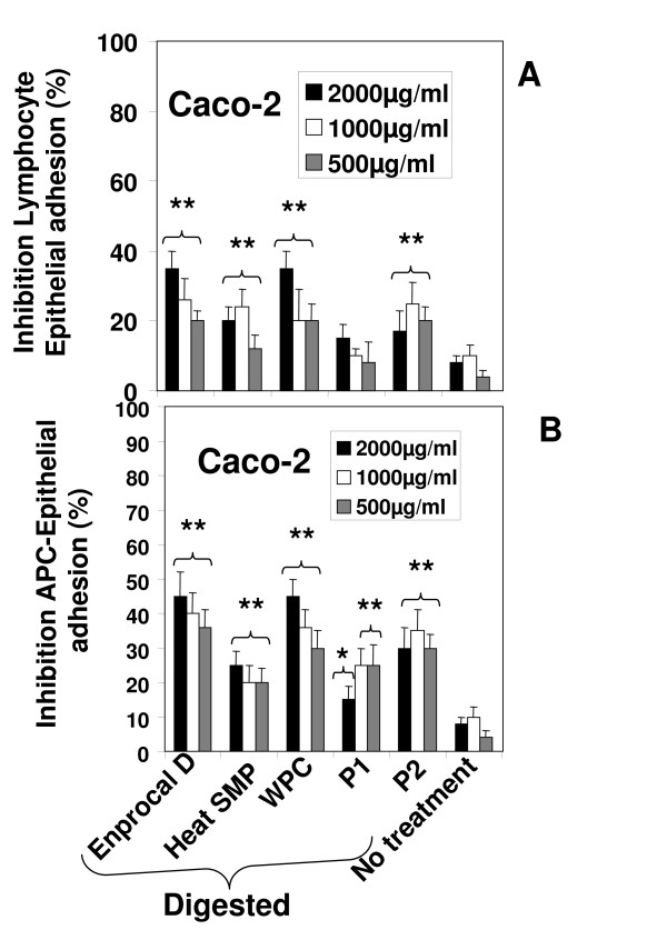
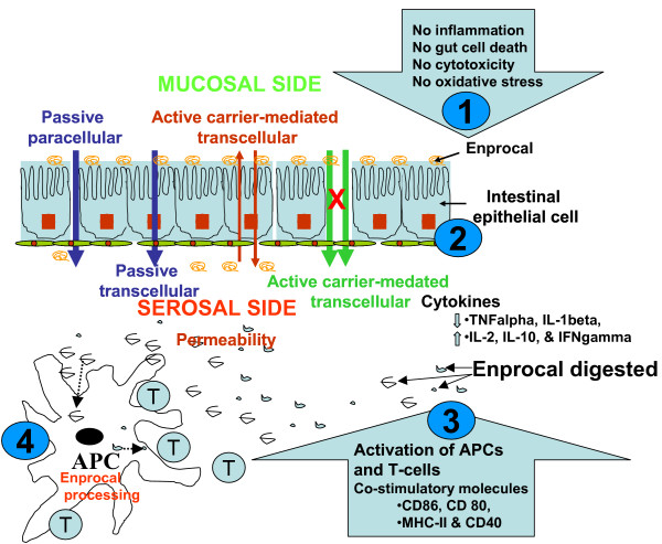
Results: Enprocal (undigested) and digested (Enprocal D) selectively enhanced cell proliferation in normal human intestinal epithelial cells (FHs74-Int) and showed no cytotoxicity. In a dose dependent manner Enprocal induced cell death in Caco-2 cells (human colon adencarcinoma epithelial cells). Digested Enprocal (Enprocal D: gut enzyme cocktail treated) maintained the intestinal integrity in transepithelial resistance (TEER) assay, increased the permeability of horseradish peroxidase (HRP) and did not induce oxidative stress to the gut epithelial cells. Enprocal D upregulated the surface expression of co-stimulatory (CD40, CD86, CD80), MHC I and MHC II molecules on PMA differentiated THP-1 macrophages in coculture transwell model, and inhibited the monocyte/lymphocyte (THP-1/Jurkat E6-1 cells)-epithelial cell adhesion. In cytokine secretion analyses, Enprocal D down-regulated the secretion of proinflammatory cytokines (IL-1beta and TNF-alpha) and up-regulated IFN-gamma, IL-2 and IL-10.
Conclusion: Our results indicate that Enprocal creates neither oxidative injury nor cytotoxicity, stimulates normal gut cell proliferation, up regulates immune cell activation markers and may aid in the production of antibodies. Furthermore, through downregulation of proinflammatory cytokines, Enprocal appears to be beneficial in reducing the effects of chronic gut inflammatory diseases such as inflammatory bowel disease (IBD). Stimulation of normal human fetal intestinal cell proliferation without cell cytotoxicity indicates it may also be given as infant food particularly for premature babies.
Figures
Similar articles
-
Development, validation and implementation of an in vitro model for the study of metabolic and immune function in normal and inflamed human colonic epithelium.Dan Med J. 2015 Jan;62(1):B4973. Dan Med J. 2015. PMID: 25557335 Review.
-
Development of an Inflammation-Triggered In Vitro "Leaky Gut" Model Using Caco-2/HT29-MTX-E12 Combined with Macrophage-like THP-1 Cells or Primary Human-Derived Macrophages.Int J Mol Sci. 2023 Apr 18;24(8):7427. doi: 10.3390/ijms24087427. Int J Mol Sci. 2023. PMID: 37108590 Free PMC article.
-
Development and assessment of an intestinal tri-cellular model to investigate the pro/anti-inflammatory potential of digested foods.Front Immunol. 2025 Feb 5;16:1545261. doi: 10.3389/fimmu.2025.1545261. eCollection 2025. Front Immunol. 2025. PMID: 39975553 Free PMC article.
-
Linoorbitides and enterolactone mitigate inflammation-induced oxidative stress and loss of intestinal epithelial barrier integrity.Int Immunopharmacol. 2018 Nov;64:42-51. doi: 10.1016/j.intimp.2018.08.012. Epub 2018 Aug 23. Int Immunopharmacol. 2018. PMID: 30145469
-
Alterations of the mucosal immune system in inflammatory bowel disease.J Gastroenterol. 1996 Dec;31(6):907-16. doi: 10.1007/BF02358624. J Gastroenterol. 1996. PMID: 9027661 Review.
Cited by
-
An optimized method for accurate quantification of cell migration using human small intestine cells.Metab Eng Commun. 2016 Mar 15;3:76-83. doi: 10.1016/j.meteno.2016.03.002. eCollection 2016 Dec. Metab Eng Commun. 2016. PMID: 29468115 Free PMC article.
-
Restored intestinal integrity, nutrients transporters, energy metabolism, antioxidative capacity and decreased harmful microbiota were associated with IUGR piglet's catch-up growth before weanling.J Anim Sci Biotechnol. 2022 Oct 14;13(1):129. doi: 10.1186/s40104-022-00770-8. J Anim Sci Biotechnol. 2022. PMID: 36229888 Free PMC article.
-
Clinical Potential of Hyperbaric Pressure-Treated Whey Protein.Healthcare (Basel). 2015 Jun 19;3(2):452-65. doi: 10.3390/healthcare3020452. Healthcare (Basel). 2015. PMID: 27417773 Free PMC article. Review.
-
Necrotizing Enterocolitis: Overview on In Vitro Models.Int J Mol Sci. 2021 Jun 23;22(13):6761. doi: 10.3390/ijms22136761. Int J Mol Sci. 2021. PMID: 34201786 Free PMC article. Review.
-
Cytoplasmic Form of Carlr lncRNA Facilitates Inflammatory Gene Expression upon NF-κB Activation.J Immunol. 2017 Jul 15;199(2):581-588. doi: 10.4049/jimmunol.1700023. Epub 2017 Jun 16. J Immunol. 2017. PMID: 28626066 Free PMC article.
References
-
- Volkert D, Berner YN, Berry E, Cederholm T, Coti BP, Milne A, Palmblad J, Schneider S, Sobotka L, Stanga Z, Lenzen-Grossimlinghaus R, Krys U, Pirlich M, Herbst B, Schütz T, Schröer W, Weinrebe W, Ockenga J, Lochs H. ESPEN Guidelines on Enteral Nutrition: Geriatrics. Clin Nutr. 2006;25:330–60. - PubMed
-
- Staples K. Nutrition Monograph Gut health. Dairy Management Inc; 2006. "Gut health and whey products"; pp. 1–12.
-
- Lasheras C, González C, García A, Patterson AM, Fernández S. Dietary intake and biochemical indicators of nutritional stress in an elderly institutionalised and non-institutionalised population. Nutr Res. 1999;19:1299–12.
-
- Kreider RB. Applications Monograph Seniors Nutrition. Dairy Management Inc; 2005. "Whey proteins and seniors nutrition"; pp. 1–8.
-
- McKay DL, Perrone G, Rasmussen H, Dallal G, Hartman W, Cao G, Prior RL, Roubenoff R, Blumberg JB. The effects of a multivitamin/mineral supplement on micronutrient status, antioxidant capacity and cytokine production in healthy older adults consuming a fortified diet. J Am Coll Nutr. 2000;19:613–621. - PubMed
Publication types
MeSH terms
Substances
LinkOut - more resources
Full Text Sources
Medical
Research Materials
