Review
. 2009 Jul;118(1):53-69.
doi: 10.1007/s00401-009-0486-3. Epub 2009 Jan 30.

Mechanisms of tau-induced neurodegeneration

Affiliations
Review

Mechanisms of tau-induced neurodegeneration

Khalid Iqbal et al. Acta Neuropathol. 2009 Jul.

Abstract

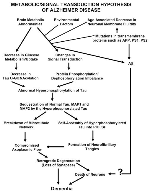
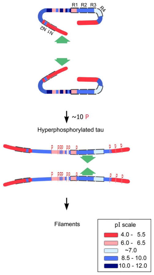
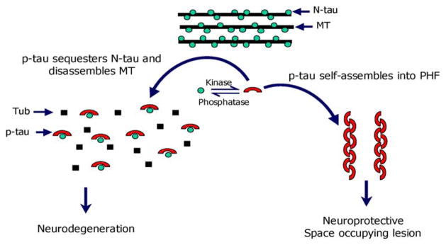
Alzheimer disease (AD) and related tauopathies are histopathologically characterized by a specific type of slow and progressive neurodegeneration, which involves the abnormal hyperphosphorylation of the microtubule associated protein (MAP) tau. This hallmark, called neurofibrillary degeneration, is seen as neurofibrillary tangles, neuropil threads, and dystrophic neurites and is apparently required for the clinical expression of AD, and in related tauopathies it leads to dementia in the absence of amyloid plaques. While normal tau promotes assembly and stabilizes microtubules, the non-fibrillized, abnormally hyperphosphorylated tau sequesters normal tau, MAP1 and MAP2, and disrupts microtubules. The abnormal hyperphosphorylation of tau, which can be generated by catalysis of several different combinations of protein kinases, also promotes its misfolding, decrease in turnover, and self-assembly into tangles of paired helical and or straight filaments. Some of the abnormally hyperphosphorylated tau ends up both amino and C-terminally truncated. Disruption of microtubules by the non-fibrillized abnormally hyperphosphorylated tau as well as its aggregation as neurofibrillary tangles probably impair axoplasmic flow and lead to slow progressive retrograde degeneration and loss of connectivity of the affected neurons. Among the phosphatases, which regulate the phosphorylation of tau, protein phosphatase-2A (PP2A), the activity of which is down-regulated in AD brain, is by far the major enzyme. The two inhibitors of PP-2A, I (1) (PP2A) and I (2) (PP2A) , which are overexpressed in AD, might be responsible for the decreased phosphatase activity. AD is multifactorial and heterogeneous and involves more than one etiopathogenic mechanism.


Figures

Fig. 1
Fig. 1
A schematic showing different major steps of the “Metabolic/Signal Transduction Hypothesis”. AD and other tauopathies require a genetic predisposition and are triggered by a variety of environmental factors, affecting one or more specific signal transduction pathways which result in a protein phosphorylation/dephosphorylation imbalance and the abnormal hyperphosphorylation of tau that leads to neurofibrillary degeneration and dementia. In AD, the protein phosphorylation/dephosphorylation imbalance in the affected neurons is generated at least in part by a decrease in the activities of tau phosphatases, i.e., PP-2A and PP-1; the activities of tau kinases such as cdk5, GSK-3, CaM kinase II and PKA might also be increased in the affected neurons. This protein phosphorylation/dephosphorylation imbalance probably involves an alteration of a specific signal transduction pathway(s) produced by an increase in the levels of an extracellular signal, e.g., FGF2 or an alteration in the molecular topology of the neuronal cell membrane or both. With age, the molecular topology of the cell membranes is altered due to a decrease in membrane fluidity. The mutations in transmembrane proteins, such as β-APP, PS1 and PS2, increase the vulnerability of the cell membrane to alteration in pathological signal transduction. The increased risk for AD in the carriers of APOE4 allele as opposed to APOE2 or APOE3 alleles might also involve alteration of signal transduction through the interaction of APOE4 with the neuronal cell membrane. Any mutation or posttranslational modification of tau that will make it a better substrate for abnormal hyperphosphorylation will also increase the risk for the disease. High cholesterol such as in Niemann Pick C disease might be involved in decreasing membrane fluidity. Decreased glucose metabolism/uptake might lead to the abnormal hyperphosphorylation of tau through a decrease in its O-GlcNA-cylation (reproduced with permission from Iqbal and Grundke-Iqbal [73])
Fig. 2
Fig. 2
A hypothetical scheme of the phosphorylation-induced self-assembly of tau. Tau self-assembles mainly through the microtubule binding domain/repeat R3 in 3R tau proteins and through R3 and R2 in 4R tau proteins (R2 and R3 have β-structure). Regions of tau molecule both N-terminal and C-terminal to the repeats are inhibitory. Hyperphosphorylation of tau neutralizes these basic inhibitory domains, enabling tau-tau interaction (phosphorylation sites indicated by red Ps). In the case of the C-terminal region beyond Pro-397 (398–441), a highly acidic segment masks the repeats. Phosphorylation (red Ps) of tau at Ser-396 and/or 404 opens this segment, allowing tau–tau interaction through the repeats. The highly basic segments and the C terminus interfere with polymerization. Upon hyperphosphorylation (phosphorylation positions indicated in red Ps), tau proteins adopt the conformation needed to polymerize into filaments (adapted from Alonso et al. [7])
Fig. 3
Fig. 3
Proposed mechanism of tau-induced neurodegeneration in AD and related tauopathies (reproduced with permission from Alonso et al. [5])

References

    1. Alafuzoff I, Iqbal K, Friden H, Adolfsson R, Winblad B. Histopathological criteria for progressive dementia disorders: clinical-pathological correlation and classification by multivariate data analysis. Acta Neuropathol. 1987;74:209–225. doi: 10.1007/BF00688184. - DOI - PubMed
    1. Alonso AD, Grundke-Iqbal I, Iqbal K. Alzheimer’s disease hyperphosphorylated tau sequesters normal tau into tangles of filaments and disassembles microtubules. Nat Med. 1996;2:783–787. doi: 10.1038/nm0796-783. - DOI - PubMed
    1. Alonso AD, Zaidi T, Grundke-Iqbal I, Iqbal K. Role of abnormally phosphorylated tau in the breakdown of microtubules in Alzheimer disease. Proc Natl Acad Sci USA. 1994;91:5562–5566. doi: 10.1073/pnas.91.12.5562. - DOI - PMC - PubMed
    1. Alonso AD, Grundke-Iqbal I, Barra HS, Iqbal K. Abnormal phosphorylation of tau and the mechanism of Alzheimer neurofibrillary degeneration: sequestration of microtubule-associated proteins 1 and 2 and the disassembly of microtubules by the abnormal tau. Proc Natl Acad Sci USA. 1997;94:298–303. doi: 10.1073/pnas.94.1.298. - DOI - PMC - PubMed
    1. Alonso AD, Li B, Grundke-Iqbal I, Iqbal K. Polymerization of hyperphosphorylated tau into filaments eliminates its inhibitory activity. Proc Natl Acad Sci USA. 2006;23:8864–8869. doi: 10.1073/pnas.0603214103. - DOI - PMC - PubMed

MeSH terms