

The expression of differentiation markers in aquaporin-3 deficient epidermis
- PMID: 19184071
- PMCID: PMC3582396
- DOI: 10.1007/s00403-009-0927-9
The expression of differentiation markers in aquaporin-3 deficient epidermis
Abstract
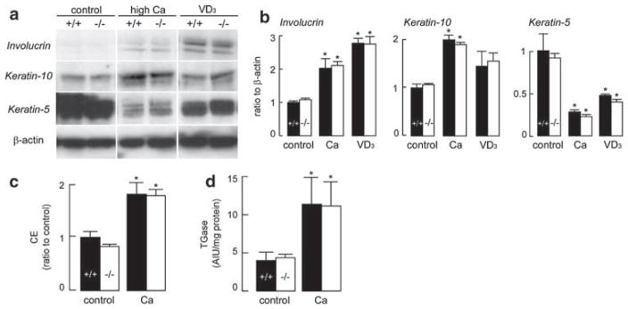
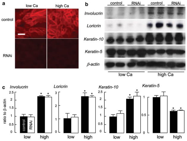
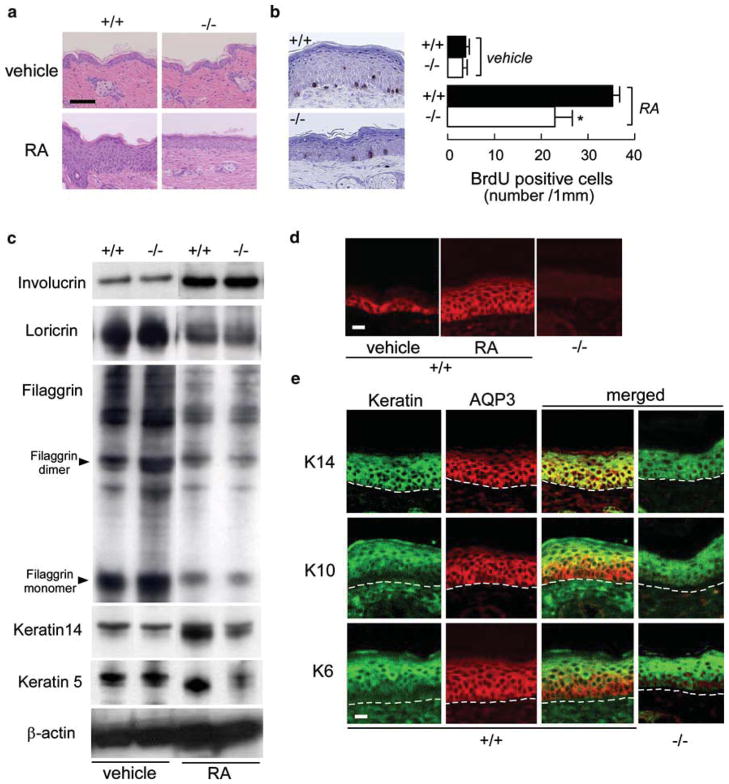
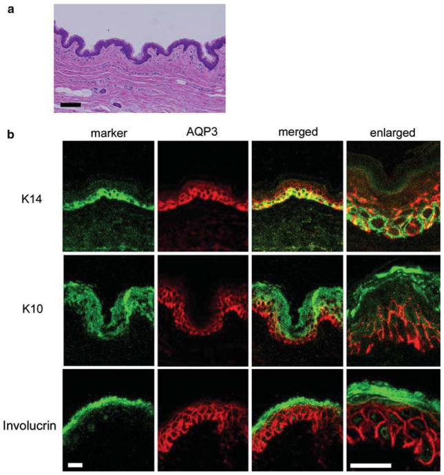
Aquaporin-3 (AQP3) is a water/glycerol transporting protein expressed strongly at the plasma membrane of keratinocytes. There is evidence for involvement of AQP3-facilitated water and glycerol transport in keratinocyte migration and proliferation, respectively. Here, we investigated the involvement of AQP3 in keratinocyte differentiation. Studies were done using AQP3 knockout mice, primary cultures of mouse keratinocytes (AQP3 knockout), neonatal human keratinocytes (AQP3 knockdown), and human skin. Cells were cultured with high Ca(2+) or 1alpha,25-dihydroxyvitamin D(3) (VD(3)) to induce differentiation. The expression of differentiation marker proteins and differentiating responses were comparable in control and AQP3-knockout or knockdown keratinocytes. Topical application of all-trans retinoic acid (RA), a known regulator of keratinocyte differentiation and proliferation, induced comparable expression of differentiation marker proteins in wildtype and AQP3 null epidermis, though with impaired RA-induced proliferation in AQP3 null mice. Immunostaining of human and mouse epidermis showed greater AQP3 expression in cells undergoing proliferation than differentiation. Our results showed little influence of AQP3 on keratinocyte differentiation, and provide further support for the proposed involvement of AQP3-facilitated cell proliferation.
Figures
Similar articles
-
Regulation of the Glycerol Transporter, Aquaporin-3, by Histone Deacetylase-3 and p53 in Keratinocytes.J Invest Dermatol. 2017 Sep;137(9):1935-1944. doi: 10.1016/j.jid.2017.04.031. Epub 2017 May 16. J Invest Dermatol. 2017. PMID: 28526298 Free PMC article.
-
Aquaporin-3 facilitates epidermal cell migration and proliferation during wound healing.J Mol Med (Berl). 2008 Feb;86(2):221-31. doi: 10.1007/s00109-007-0272-4. Epub 2007 Oct 30. J Mol Med (Berl). 2008. PMID: 17968524
-
PPARgamma activators stimulate aquaporin 3 expression in keratinocytes/epidermis.Exp Dermatol. 2011 Jul;20(7):595-9. doi: 10.1111/j.1600-0625.2011.01269.x. Epub 2011 Apr 4. Exp Dermatol. 2011. PMID: 21457357 Free PMC article.
-
Roles of aquaporin-3 in the epidermis.J Invest Dermatol. 2008 Sep;128(9):2145-51. doi: 10.1038/jid.2008.70. Epub 2008 Jun 12. J Invest Dermatol. 2008. PMID: 18548108 Review.
-
Aquaporin-3 in the epidermis: more than skin deep.Am J Physiol Cell Physiol. 2020 Jun 1;318(6):C1144-C1153. doi: 10.1152/ajpcell.00075.2020. Epub 2020 Apr 8. Am J Physiol Cell Physiol. 2020. PMID: 32267715 Free PMC article. Review.
Cited by
-
Aquaporin-3 re-expression induces differentiation in a phospholipase D2-dependent manner in aquaporin-3-knockout mouse keratinocytes.J Invest Dermatol. 2015 Feb;135(2):499-507. doi: 10.1038/jid.2014.412. Epub 2014 Sep 18. J Invest Dermatol. 2015. PMID: 25233074 Free PMC article.
-
Increased differentiation capacity of bone marrow-derived mesenchymal stem cells in aquaporin-5 deficiency.Stem Cells Dev. 2012 Sep 1;21(13):2495-507. doi: 10.1089/scd.2011.0597. Epub 2012 Apr 20. Stem Cells Dev. 2012. PMID: 22420587 Free PMC article.
-
An aquaporin 3-notch1 axis in keratinocyte differentiation and inflammation.PLoS One. 2013 Nov 8;8(11):e80179. doi: 10.1371/journal.pone.0080179. eCollection 2013. PLoS One. 2013. PMID: 24260356 Free PMC article.
-
Analysis of aquaporin 9 expression in human epidermis and cultured keratinocytes.FEBS Open Bio. 2014 Jun 26;4:611-6. doi: 10.1016/j.fob.2014.06.004. eCollection 2014. FEBS Open Bio. 2014. PMID: 25161869 Free PMC article.
-
Regulation of the Glycerol Transporter, Aquaporin-3, by Histone Deacetylase-3 and p53 in Keratinocytes.J Invest Dermatol. 2017 Sep;137(9):1935-1944. doi: 10.1016/j.jid.2017.04.031. Epub 2017 May 16. J Invest Dermatol. 2017. PMID: 28526298 Free PMC article.
References
-
- Bellemère G, Von Stetten O, Oddos T. Retinoic acid increases aquaporin 3 expression in normal human skin. J Invest Dermatol. 2008;128:542–548. - PubMed
-
- Eckert RL, Crish JF, Robinson NA. The epidermal keratinocyte as a model for the study of gene regulation and cell differentiation. Physiol Rev. 1997;77:397–424. - PubMed
Publication types
MeSH terms
Substances
Grants and funding
LinkOut - more resources
Full Text Sources
Other Literature Sources
Miscellaneous
