. 2009 Mar;49(3):871-9.
doi: 10.1002/hep.22736.

Apotopes and the biliary specificity of primary biliary cirrhosis

Affiliations

Apotopes and the biliary specificity of primary biliary cirrhosis

Ana Lleo et al. Hepatology. 2009 Mar.

Abstract

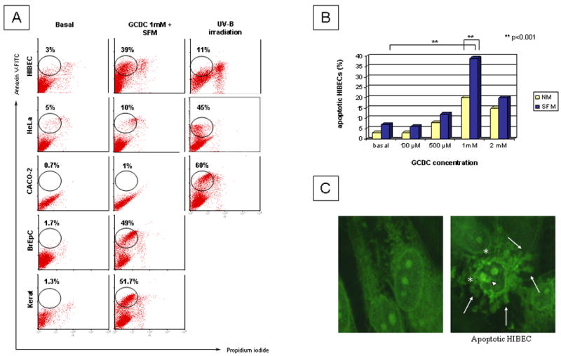
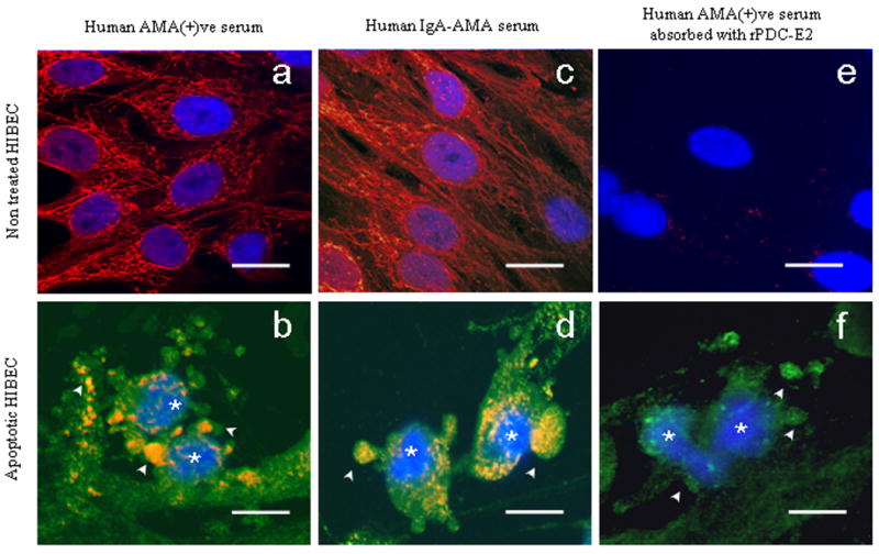
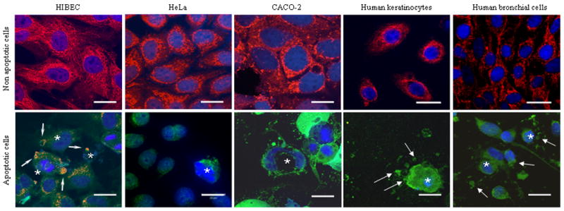
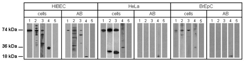
Primary biliary cirrhosis (PBC) is characterized by antimitochondrial antibodies (AMAs), directed to the E2 component of the pyruvate dehydrogenase complex (PDC-E2). Notwithstanding the presence of mitochondria in virtually all nucleated cells, the destruction in PBC is limited to small intrahepatic bile ducts. The reasons for this tissue specificity remain unknown, although biliary epithelial cells (BECs) uniquely preserve the PDC-E2 epitope following apoptosis. Notably, PBC recurs in an allogeneic transplanted liver, suggesting generic rather than host PBC-specific susceptibility of BEC. We used cultured human intrahepatic BECs (HIBECs) and other well-characterized cell lines, including, HeLa, CaCo-2 cells, and nontransformed human keratinocytes and bronchial epithelial cells, to determine the integrity and specific localization of PDC-E2 during induced apoptosis. All cell lines, both before and after apoptosis, were tested with sera from patients with PBC (n = 30), other autoimmune liver and rheumatic diseases (n = 20), and healthy individuals (n = 20) as well as with a mouse monoclonal antibody against PDC-E2 and AMA with an immunoglobulin A isotype. PDC-E2 was found to localize unmodified within apoptotic blebs of HIBECs, but not within blebs of various other cell lineages studied. The fact that AMA-containing sera reacted with PDC-E2 on apoptotic BECs without a requirement for permeabilization suggests that the autoantigen is accessible to the immune system during apoptosis.

Conclusion: Our data indicate that the tissue (cholangiocyte) specificity of the autoimmune injury in PBC is a consequence of the unique characteristics of HIBECs during apoptosis and can be explained by exposure to the immune system of intact immunoreactive PDC-E2 within apoptotic blebs.


Figures

Figure 1
Figure 1
Human intrahepatic biliary epithelial cells (HIBEC) and four other cell types were cultured in normal media (NM) or serum free media (SFM) under optimal conditions to induce apoptosis, either by exposure to bile salts (glycochenodeoxycholate, GCDC) or ultraviolet B (UV-B) irradiation (see Methods). A. Induction of apoptosis was investigated by flow cytometry after double staining with FITC-labeled Annexin V and propidium iodide. The use of 1 mM GCDC in the absence of serum, or growth factors, induced apoptosis in 39% of HIBEC, 49% of BrEpC and 51.7% of keratinocytes whereas no significant apoptotic response was observed in transformed cell lines. UV-B irradiation induced apoptosis in just 11% of HIBEC and a higher level of double positive PI/AnnexinV cells, most probably referable to necrotic cells. B. Different concentrations of GCDC in NM or SFM were tested to establish pro-apoptotic concentrations for HIBECs, with 1mM proving optimal. C. HIBEC after exposure to GCDC 1mM in SFM were assessed by confocal microscopy, revealing typical apoptotic morphology with blebbing of the membrane (*), nucleolar degeneration (short arrow) and apoptotic fragments (long arrow). 100 × oil immersion objective was used.
Figure 2
Figure 2
Staining of non-apoptotic or apoptotic HIBEC using an AMA-positive PBC serum (panels a, b), serum from a PBC patient with monoclonal production of IgA-AMA (panels c, d), and using the same AMA-positive serum after absorbtion with recombinant PDC-E2 (panels e, f). The immunofluorescence staining was performed with three fluorocromes: Cy3-conjugated secondary antibody (red, yellow-orange when co-stained with green), FITC-labeled Annexin-V (green), and DAPI (blue) for apoptosis detection. Apoptotic cells (*) were identified by morphological criteria: high nuclear density, chromatin condensation and nuclear fragmentation revealed with DAPI (blue), and characteristic blebbing of the cell membrane revealed with Annexin-V (green). Positive staining of blebs and apoptotic fragments (arrows) was observed using unabsorbed PBC sera and was virtually absent after absorption. Apoptosis was confirmed in all experiments (*). Scale bar represents 20 μm.
Figure 3
Figure 3
Apoptosis-dependent anti-PDC-E2 staining in various human cell types, with non-apoptotic cells shown in upper row and apoptotic cells in lower row. Immunofluorescence staining was performed with three fluorochromes, mAb against PDC-E2 and Cy3-conjugated secondary antibody (red, yellow-orange when co-stained with green); FITC-labeled Annexin-V (green); and DAPI (blue) for apoptosis detection. Apoptotic cells (*) were identified as described in Figure 2. Non- apoptotic cells (upper row) have a normal AMA pattern of immunofluorescence staining (red). Apoptotic cells (lower row) show differences between HIBEC that retained mitochondrial staining within blebs and fragments (arrow) after apoptosis, and other cell types (CACO-2 cells, HeLa cells, human keratinocytes and human bronchial epithelial cells) that lost mitochondrial staining after apoptosis. 100× oil immersion objective. Scale bar represents 20 μm.
Figure 4
Figure 4
Western blots showing that PDC-E2 is localized unmodified within apoptotic bodies (AB) from human intrahepatic biliary cells (HIBEC) but not from other cell lineages. ABs were isolated by filtration and ultracentrifugation after induction of apoptosis (see Methods). PDC-E2 was detected by Western blot in lysates obtained from non-apoptotic cells and AB using (1) mAb against PDC-E2, (2) AMA positive serum, (3) AMA-IgA isotype, (4) mAb against caspase 3, and (5) serum from a healthy control. PDC-E2 was detected in ABs from HIBEC when tested with monoclonal Ab and with PBC sera of two different Ig isotypes. ABs from HeLa cells and BrEpC did not show the presence of PDC-E2 whereas, as expected, lysates from non-apoptotic cells did so. The 18 kDa subunit of caspase-3 generated during apoptosis is localized within apoptotic blebs, and hence exposure to mAb to caspase-3 was used to validate formation of blebs.
Figure 5
Figure 5
Staining of non-apoptotic (upper row) and apoptotic (lower row) HIBEC in absence or presence of a membrane permeabilizing agent, Triton X-100. AMA staining in normal cells requires pemeabilization of the cell (a); lacking this, no staining is observed due to retention of membrane integrity (c); apoptotic HIBEC (*) stained with a mAb against PDC-E2, with positive staining demonstrable within the blebs (arrow) (b); apoptotic HIBEC retained the bleb staining even without permeabilization (d). No staining was observed with serum from patients with AMA negative PBC, PSC, AIH, SLE, CHC or healthy subjects (not shown). Scale bar represents 20 μm.

References

    1. Savill J, Dransfield I, Gregory C, Haslett C. A blast from the past: clearance of apoptotic cells regulates immune responses. Nat Rev Immunol. 2002;2:965–975. - PubMed
    1. Ravichandran KS, Lorenz U. Engulfment of apoptotic cells: signals for a good meal. Nat Rev Immunol. 2007;7:964–974. - PubMed
    1. Torok NJ. Apoptotic cell death takes its toll. Hepatology. 2007;46:1323–1325. - PubMed
    1. Perniok A, Wedekind F, Herrmann M, Specker C, Schneider M. High levels of circulating early apoptic peripheral blood mononuclear cells in systemic lupus erythematosus. Lupus. 1998;7:113–118. - PubMed
    1. Ruiz-Arguelles A, Brito GJ, Reyes-Izquierdo P, Perez-Romano B, Sanchez-Sosa S. Apoptosis of melanocytes in vitiligo results from antibody penetration. J Autoimmun. 2007;29:281–286. - PubMed

Publication types

MeSH terms