

Enhanced estrogen-induced proliferation in obese rat endometrium
- PMID: 19185100
- PMCID: PMC2880878
- DOI: 10.1016/j.ajog.2008.08.064
Enhanced estrogen-induced proliferation in obese rat endometrium
Abstract
Objective: We tested the hypothesis that the proliferative estrogen effect on the endometrium is enhanced in obese vs lean animals.
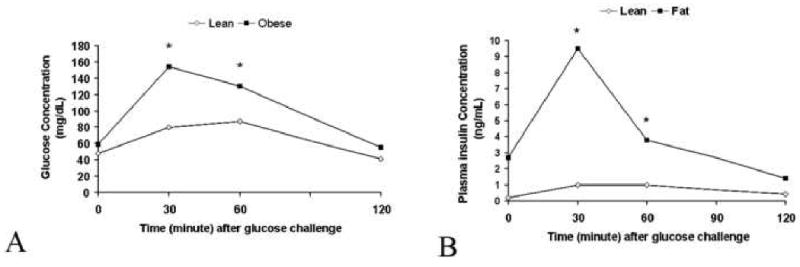
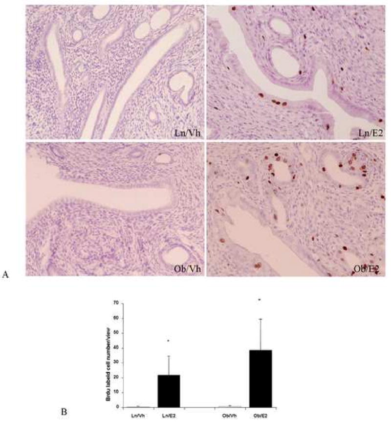
Study design: Using Zucker fa/fa obese rats and lean control, we examined endometrial cell proliferation and the expression patterns of certain estrogen-regulated proproliferative and antiproliferative genes after short-term treatment with estradiol.
Results: No significant morphologic/histologic difference was seen between the obese rats and the lean rats. Estrogen-induced proproliferative genes cyclin A and c-Myc messenger RNA expression were significantly higher in the endometrium of obese rats compared with those of the lean control. Expression of the antiproliferative gene p27Kip1 was suppressed by estrogen treatment in both obese and lean rats; however, the decrease was more pronounced in obese rats. Estrogen more strongly induced the antiproliferative genes retinaldehyde dehydrogenases 2 and secreted frizzled-related protein 4 in lean rats but had little or no effect in obese rats.
Conclusion: Enhancement of estrogen-induced endometrial proproliferative gene expression and suppression of antiproliferative gene expression was seen in the endometrium of obese vs lean animals.
Figures


Similar articles
-
Chemopreventive effects of metformin on obesity-associated endometrial proliferation.Am J Obstet Gynecol. 2013 Jul;209(1):24.e1-24.e12. doi: 10.1016/j.ajog.2013.03.008. Epub 2013 Mar 15. Am J Obstet Gynecol. 2013. PMID: 23500454 Free PMC article.
-
CGRRF1 as a novel biomarker of tissue response to metformin in the context of obesity.Gynecol Oncol. 2014 Apr;133(1):83-9. doi: 10.1016/j.ygyno.2013.12.006. Gynecol Oncol. 2014. PMID: 24680596 Free PMC article.
-
[Effects of metformin on the estrogen-induced proliferation and the expression of ER in human endometrial cancer cells].Zhonghua Fu Chan Ke Za Zhi. 2014 Dec;49(12):932-7. Zhonghua Fu Chan Ke Za Zhi. 2014. PMID: 25608995 Chinese.
-
Estrogen and the endometrium: lessons learned from gene expression profiling in rodents and human.Hum Reprod Update. 2007 Jul-Aug;13(4):405-17. doi: 10.1093/humupd/dmm009. Hum Reprod Update. 2007. PMID: 17584823 Review.
-
Hormonal regulation and localization of estrogen, progestin and androgen receptors in the endometrium of nonhuman primates: effects of progesterone receptor antagonists.Arch Histol Cytol. 2004 Dec;67(5):393-409. doi: 10.1679/aohc.67.393. Arch Histol Cytol. 2004. PMID: 15781981 Review.
Cited by
-
Kingiodendron pinnatum, a pharmacologically effective alternative for Saraca asoca in an Ayurvedic preparation, Asokarishta.J Tradit Complement Med. 2017 Jun 26;8(1):244-250. doi: 10.1016/j.jtcme.2017.06.005. eCollection 2018 Jan. J Tradit Complement Med. 2017. PMID: 29322015 Free PMC article.
-
Associations Between Endometriosis and Gut Microbiota.Reprod Sci. 2021 Aug;28(8):2367-2377. doi: 10.1007/s43032-021-00506-5. Epub 2021 Mar 3. Reprod Sci. 2021. PMID: 33660232 Free PMC article.
-
Metformin as a Potential Treatment Option for Endometriosis.Cancers (Basel). 2022 Jan 24;14(3):577. doi: 10.3390/cancers14030577. Cancers (Basel). 2022. PMID: 35158846 Free PMC article. Review.
-
Skeletal and Uterotrophic Effects of Endoxifen in Female Rats.Endocrinology. 2017 Oct 1;158(10):3354-3368. doi: 10.1210/en.2016-1871. Endocrinology. 2017. PMID: 28977607 Free PMC article.
-
Mesenchymal stem cells from adipose tissue prone to lose their stemness associated markers in obesity related stress conditions.Sci Rep. 2024 Aug 24;14(1):19702. doi: 10.1038/s41598-024-70127-w. Sci Rep. 2024. PMID: 39181924 Free PMC article.
References
-
- McCourt CK, Mutch DG, Gibb RK, et al. Body mass index: relationship to clinical, pathologic and features of microsatellite instability in endometria cancer. Gynecol Oncol. 2007;104:535–9. - PubMed
-
- Schottenfeld D. Epidemiology of endometrial neoplasia. J Cell Biochem. 1995 23:151–9. - PubMed
-
- Pettersson B, Bergstrom R, Johansson ED. Serum estrogens and androgens in women with endometrial carcinoma. Gynecol Oncol. 1986;25:223–33. - PubMed
-
- Potischman N, Hoover RN, Brinton LA, et al. Case-control study of endogenous steroid hormones and endometrial cancer. J Natl Cancer Inst. 1996;88:1127–35. - PubMed
-
- Friberg E, Mantzoros CS, Wolk A. Diabetes and risk of endometrial cancer: a population-based prospective cohort study. Cancer Epidemiol Biomarkers Prev. 2007;16:276–80. - PubMed
Publication types
MeSH terms
Substances
Grants and funding
LinkOut - more resources
Full Text Sources
Medical
