

Notch signaling and Hes labeling in the normal and drug-damaged organ of Corti
- PMID: 19185606
- PMCID: PMC2796274
- DOI: 10.1016/j.heares.2008.12.008
Notch signaling and Hes labeling in the normal and drug-damaged organ of Corti
Abstract
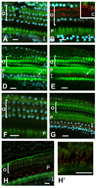
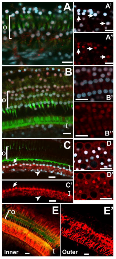
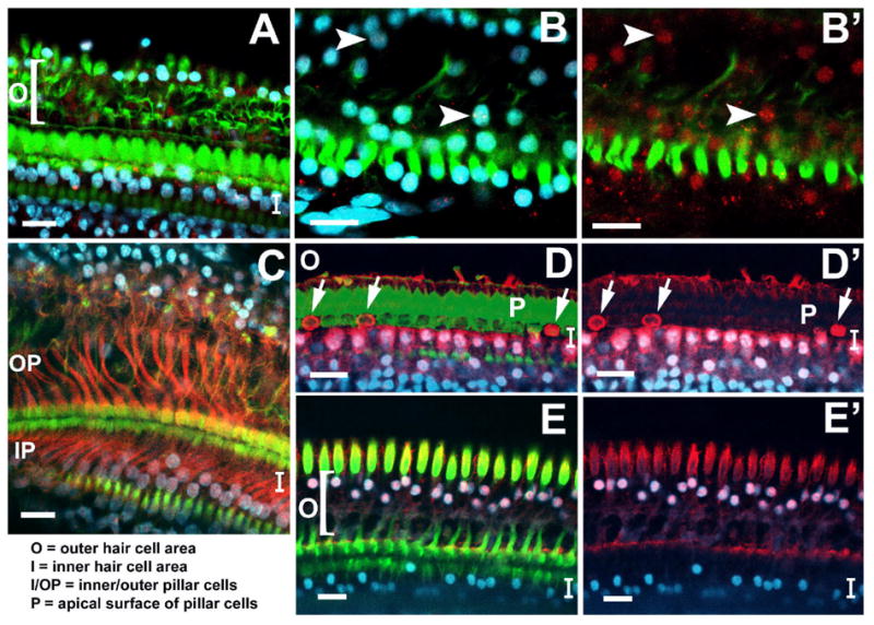
During the development of the inner ear, the Notch cell signaling pathway is responsible for the specification of the pro-sensory domain and influences cell fate decisions. It is assumed that Notch signaling ends during maturity and cannot be reinitiated to alter the fate of new or existing cells in the organ of Corti. This is in contrast to non-mammalian species which reinitiate Delta 1-Notch1 signaling in response to trauma in the auditory epithelium, resulting in hair cell regeneration through transdifferentiation and/or mitosis. We report immunohistochemical data and Western protein analysis showing that in the aminoglycoside-damaged guinea pig organ of Corti, there is an increase in proteins involved in Notch activation occurring within 24h of a chemical hair cell lesion. The signaling response is characterized by the increased presence of Jagged1 ligand in pillar and Deiters cells, Notch1 signal in surviving supporting cell nuclei, and the absence of Jagged2 and Delta-like1. The pro-sensory bHLH protein Atoh1 was absent at all time points following an ototoxic lesion, while the repressor bHLH transcription factors Hes1 and Hes5 were detected in surviving supporting cell nuclei in the former inner and outer hair cell areas, respectively. Notch pathway proteins peaked at 2 weeks, decreased at 1 month, and nearly disappeared by 2 months. These results indicate that the mammalian auditory epithelium retains the ability to regulate Notch signaling and Notch-dependent Hes activity in response to cellular trauma and that the signaling is transient. Additionally, since Hes activity antagonizes the transcription of pro-sensory Atoh1, the presence of Hes after a lesion may prohibit the occurrence of transdifferentiation in the surviving supporting cells.
Figures

References
-
- Abrashkin KA, Izumikawa M, Miyazawa T, Wang CH, Crumling MA, Swiderski DL, Beyer LA, Gong TW, Raphael Y. The fate of outer hair cells after acoustic or ototoxic insults. Hear Res 2006 - PubMed
-
- Adler HJ, Raphael Y. New hair cells arise from supporting cell conversion in the acoustically damaged chick inner ear [published erratum appears in Neurosci Lett 1996 May 24;210(1):73] Neurosci Lett. 1996;205:17–20. - PubMed
-
- Baird RA, Steyger PS, Schuff NR. Mitotic and nonmitotic hair cell regeneration in the bullfrog vestibular otolith organs. Ann N Y Acad Sci. 1996;781:59–70. - PubMed
-
- Bermingham-McDonogh O, Rubel EW. Hair cell regeneration: winging our way towards a sound future. Curr Opin Neurobiol. 2003;13:119–26. - PubMed
-
- Bray SJ. Notch signalling: a simple pathway becomes complex. Nat Rev Mol Cell Biol. 2006;7:678–89. - PubMed
Publication types
MeSH terms
Substances
Grants and funding
LinkOut - more resources
Full Text Sources
Other Literature Sources
