

Review
doi: 10.1016/j.ceb.2009.01.006.
Epub 2009 Jan 29.
Actin and endocytosis: mechanisms and phylogeny
Affiliations
- PMID: 19186047
- PMCID: PMC2670849
- DOI: 10.1016/j.ceb.2009.01.006
Item in Clipboard
Review
Actin and endocytosis: mechanisms and phylogeny
Curr Opin Cell Biol.
2009 Feb.
Abstract
The regulated assembly of actin filament networks is a crucial part of endocytosis, with crucial temporal and spatial relationships between proteins of the endocytic and actin assembly machinery. Of particular importance has been a wealth of studies in budding and fission yeast. Cell biology approaches, combined with molecular genetics, have begun to uncover the complexity of the regulation of actin dynamics during the endocytic process. In a wide range of organisms, clathrin-mediated endocytosis appears to be linked to Arp2/3-mediated actin assembly. The conservation of the components, across a wide range eukaryotic species, suggests that the partnership between endocytosis and actin may be evolutionarily ancient.
Figures
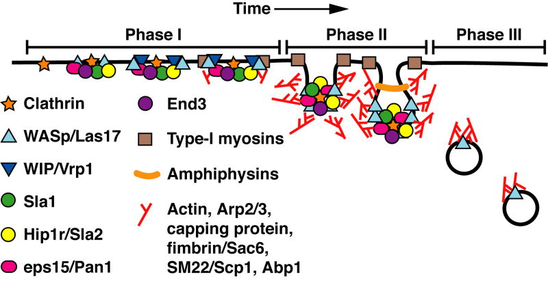
Model of actin patch assembly and movement during endocytosis in S. cerevisiae. The phases of patch movement we have defined and described in the text are overlaid on the model. This model is derived from the results of numerous works described and referenced herein.
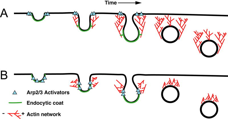
Models of actin assembly during the invagination of the endocytic membrane. These models are derived from the results of numerous works described and referenced herein. The orientation of the actin filaments is indicated in the legend with a “+” for barbed ends and a “-” for pointed ends. Model A. The site of endocytosis is initially marked by recruitment of endocytic coat proteins and Arp2/3 activator proteins. The Arp2/3 activator proteins recruit Arp2/3 to nucleate an actin network from the plasma membrane. The network grows from these sites, with new actin monomers being added adjacent to the plasma membrane and the older parts of the network flowing into the cytoplasm. The endocytic coat proteins are anchored to this network, such that the flow of this network pulls the endocytic membrane into the cell. Model B. As in (A), sites of endocytosis are marked by recruitment of endocytic coat proteins and Arp2/3 activators. Immuno-EM studies suggest that the initial curvature of the membrane may occur prior to actin polymerization (see text). An actin network is nucleated from this invagination. The force of polymerization squeezes against this invagination, helping drive the endocytic coat into the cytoplasm, as well as providing lateral force for vesicle scission. Once scission occurs, the actin filaments are asymmetrically arranged around the endocytic vesicle and can propel its movement through the cytoplasm.
References
-
- Ayscough KR. Coupling actin dynamics to the endocytic process in Saccharomyces cerevisiae. Protoplasma. 2005;22:681–88. - PubMed
-
- Kaksonen M, Toret CP, Drubin DG. Harnessing actin dynamics for clathrin-mediated endocytosis. Nat Rev Mol Cell Biol. 2006;7:404–414. - PubMed
-
- Smythe E, Ayscough KR. Actin regulation in endocytosis. J Cell Sci. 2006;119:4589–4598. - PubMed
Publication types
MeSH terms
Substances
Grants and funding
LinkOut - more resources
Full Text Sources
Molecular Biology Databases
