. 2009 Feb;96(3):1151-8.
doi: 10.1016/j.bpj.2008.10.030.

UVA generates pyrimidine dimers in DNA directly

Affiliations

UVA generates pyrimidine dimers in DNA directly

Yong Jiang et al. Biophys J. 2009 Feb.

Abstract

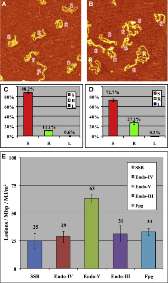
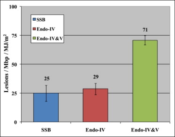
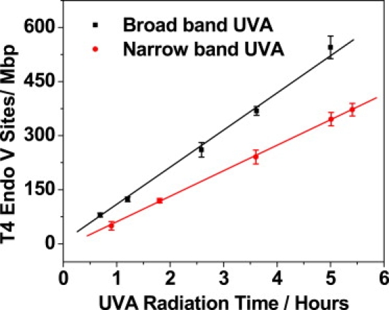
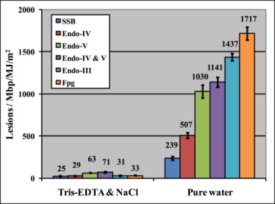
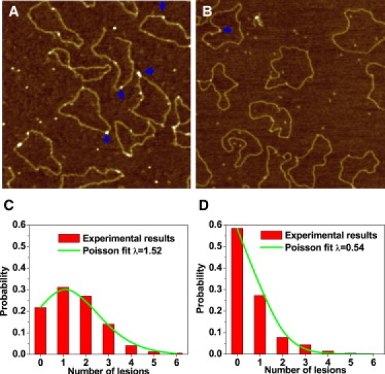
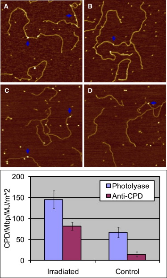
There is increasing evidence that UVA radiation, which makes up approximately 95% of the solar UV light reaching the Earth's surface and is also commonly used for cosmetic purposes, is genotoxic. However, in contrast to UVC and UVB, the mechanisms by which UVA produces various DNA lesions are still unclear. In addition, the relative amounts of various types of UVA lesions and their mutagenic significance are also a subject of debate. Here, we exploit atomic force microscopy (AFM) imaging of individual DNA molecules, alone and in complexes with a suite of DNA repair enzymes and antibodies, to directly quantify UVA damage and reexamine its basic mechanisms at a single-molecule level. By combining the activity of endonuclease IV and T4 endonuclease V on highly purified and UVA-irradiated pUC18 plasmids, we show by direct AFM imaging that UVA produces a significant amount of abasic sites and cyclobutane pyrimidine dimers (CPDs). However, we find that only approximately 60% of the T4 endonuclease V-sensitive sites, which are commonly counted as CPDs, are true CPDs; the other 40% are abasic sites. Most importantly, our results obtained by AFM imaging of highly purified native and synthetic DNA using T4 endonuclease V, photolyase, and anti-CPD antibodies strongly suggest that CPDs are produced by UVA directly. Thus, our observations contradict the predominant view that as-yet-unidentified photosensitizers are required to transfer the energy of UVA to DNA to produce CPDs. Our results may help to resolve the long-standing controversy about the origin of UVA-produced CPDs in DNA.


Figures

Figure 1
Figure 1
AFM images on APS-mica (48) of different pUC18 DNAs that were subjected to 1.3 MJ/m2 UVA radiation and different enzyme treatments before imaging. DNA was dialyzed in 10 mM Tris-HCl, 1 mM EDTA, and 100 mM NaCl buffer, and irradiated in the same solution by UVA. After that, the sample was diluted back to a suitable buffer for different enzyme incubations: (A) no enzyme treatment as control, (B) T4 endonuclease V. The scan size in all the images is 1 × 1 μm2. (C and D) Histograms of the occurrence of various configurations of pUC18 plasmids determined from the AFM images, such as these shown in A and B. Color code: red, supercoiled DNA (S); green, relaxed circular plasmids (R); blue, linear DNA (L). The error bars in the figures represent the SD. Each histogram is based on 600–1000 DNA molecules from 30–36 AFM images. (E) Histogram summarizes the number of different lesions/Mbp/MJ/m2 after UVA irradiation and specific enzyme treatments. The values shown in the histogram represent averages from two to five separate experiments.
Figure 2
Figure 2
Histogram summarizes the actual CPD number/Mbp/MJ/m2 of UVA-irradiated DNA by combining E. coli endonuclease IV and T4 endonuclease V enzyme treatments. The values shown in the histogram represent averages from two to five separate experiments.
Figure 3
Figure 3
T4 endonuclease V-sensitive sites generated by broadband (without filter) and narrowband (with filter) UVA irradiation as a function of radiation time.
Figure 4
Figure 4
Histograms show the different lesions after UVA irradiation in 10 mM Tris-HCl, 1 mM EDTA, and 100 mM NaCl, and ultrapure Millipore water. DNA was treated by different enzymes at their preferred buffer condition and then diluted by regular imaging buffer (10 mM Tris-HCl, 1 mM EDTA, and 100 mM NaCl) before deposition onto APS-mica.
Figure 5
Figure 5
AFM images show photolyases binding to the CPD sites of pUC18 (some of them marked by blue arrows) with (A) 1 MJ/m2 UVA radiation and (B) no UVA radiation. Irradiation was performed on dialyzed plasmids suspended in pure water. The scan size in all the images is 1 × 1 μm2. (C and D) Histograms show the distribution of photolyase on pUC18 molecules as shown in A and B. The curves show the Poisson distribution fits, which give the average damage λ = 1.52/plasmid for the UVA-radiated DNA and 0.54 for control DNA, respectively.
Figure 6
Figure 6
(A and B) AFM images show photolyases binding to the CPD sites of poly(dA)-poly(dT) with (A) 6 MJ/m2 UVA radiation, and (B) no UVA radiation. (C and D) AFM images show antithymine-dimer antibodies binding to the CPD sites of poly(dA)-poly(dT) with (C) 6 MJ/m2 UVA radiation, and (D) no UVA radiation as control. The scan size in all the images is 1 × 1 μm2. The histograms compare the lesions detected by photolyase and antithymine-dimer antibody on UVA-irradiated poly(dA)-poly(dT) and intact poly(dA)-poly(dT).

References

    1. Setlow R.B. Wavelengths in sunlight effective in producing skin cancer—theoretical analysis. Proc. Natl. Acad. Sci. USA. 1974;71:3363–3366. - PMC - PubMed
    1. Morrison H. John Wiley & Sons; New York: 1990. Bioorganic Photochemistry, Vol. 1, Photochemistry and the Nucleic Acids.
    1. Friedberg E.C., Walker G.C., Siede W. ASM Press; Washington, D.C.: 1995. DNA Repair and Mutagenesis.
    1. Sinha R.P., Hader D.P. UV-induced DNA damage and repair: a review. Photochem. Photobiol. Sci. 2002;1:225–236. - PubMed
    1. Cadet J., Sage E., Douki T. Ultraviolet radiation-mediated damage to cellular DNA. Mutat. Res. 2005;571:3–17. - PubMed

Publication types

MeSH terms