. 2009 Jan 29;61(2):213-9.
doi: 10.1016/j.neuron.2008.11.024.

Astrocytic modulation of sleep homeostasis and cognitive consequences of sleep loss

Affiliations

Astrocytic modulation of sleep homeostasis and cognitive consequences of sleep loss

Michael M Halassa et al. Neuron. .

Abstract

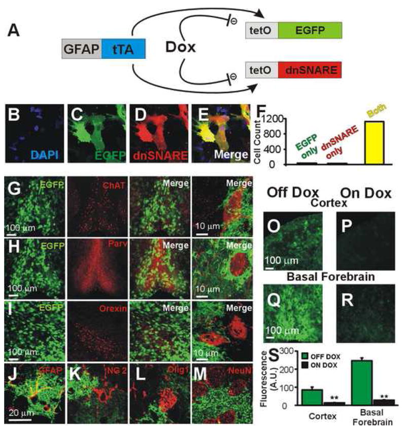
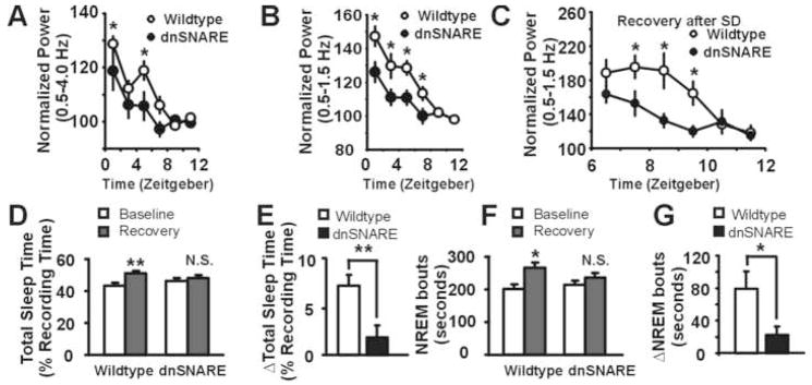
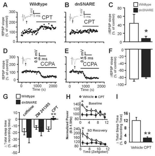
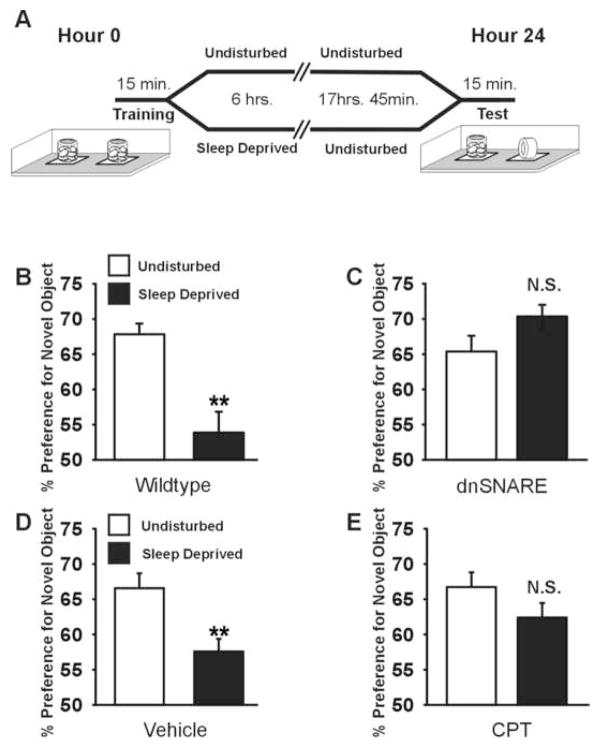
Astrocytes modulate neuronal activity by releasing chemical transmitters via a process termed gliotransmission. The role of this process in the control of behavior is unknown. Since one outcome of SNARE-dependent gliotransmission is the regulation of extracellular adenosine and because adenosine promotes sleep, we genetically inhibited the release of gliotransmitters and asked if astrocytes play an unsuspected role in sleep regulation. Inhibiting gliotransmission attenuated the accumulation of sleep pressure, assessed by measuring the slow wave activity of the EEG during NREM sleep, and prevented cognitive deficits associated with sleep loss. Since the sleep-suppressing effects of the A1 receptor antagonist CPT were prevented following inhibition of gliotransmission and because intracerebroventricular delivery of CPT to wild-type mice mimicked the transgenic phenotype, we conclude that astrocytes modulate the accumulation of sleep pressure and its cognitive consequences through a pathway involving A1 receptors.


Figures

Figure 1
Figure 1. Conditional, astrocytes-specific expression of dnSNARE in brain regions involved in sleep regulation
(A) Cartoon depicting GFAP promoter driving the expression of dnSNARE and EGFP (reporter) in astrocytes. Dox suppresses expression of both transgenes. (B–F) EGFP is a reliable marker for the dnSNARE, as 97.3% of cultured astrocytes expressing the soluble SNARE domain of Synaptobrevin II (dnSNARE) are EGFP+. In brain sections, astrocytes expressing transgenes (EGFP; green) are in proximity to cholinergic, choline acetyl transferase (ChAT) neurons (red, G) and to noncholinergic, parvalbumin (Parv) positive neurons (red, H) in the basal forebrain and surround orexinergic neurons, (red, I), in the lateral hypothalamus. (J–M) Single optical sections showing that EGFP colocalizes with the astrocytic marker GFAP (J) but not with markers of other glia (NG2, K; Olig1, L) or neurons (NeuN, M). Calibrations for K–M correspond to J. Placing dnSNARE mice on a Dox-containing diet suppresses transgene expression in the cortex (O, P) and the basal forebrain (Q, R). (S) Quantification of O–R (n = 3 animals in each group, **, p < 0.001, unpaired t-test). Differences in EGFP expression between cortex and basal forebrain correlate with relative levels of GFAP expression in these brain regions.
Figure 2
Figure 2. Gliotransmission is essential for sleep pressure accumulation
(A) SWA (0.5–4.0 Hz) during NREM sleep across the light phase is decreased in the dnSNARE animals (n = 7 animals) compared to their wildtype littermates (n = 8 animals). (ANOVA, p<0.002, F=10.413, posthoc test, *, p < 0.05). (B) low-frequency SWA (0.5–1.5Hz) is reduced across the light phase in the dnSNARE compared to wildtype animals. (ANOVA, p<0.001, F=21.247, posthoc test, *, p <0.05). (C) Following sleep deprivation (SD), low frequency SWA is decreased in the dnSNARE animals (ANOVA, p<0.001, F=7.911, posthoc test, *, p < 0.05). (D) Sleep deprivation increases TST in wildtype (n = 9) but not dnSNARE animals (n = 8) during an 18 hour recovery period compared to a baseline period **, p < 0.001. (E) The increase in total sleep time (TST) after SD over the 18 hours of recovery in the dnSNARE is blunted when directly compared to wildtype animals (unpaired t-test, **, p < 0.01. N.S., non-significant).. (E) Sleep deprivation causes an increase in NREM bout durations in the subsequent 18-hours of recovery in the wildtype animals *, p < 0.05. The increase is not statistically significant in the dnSNARE. (G) When compared directly, the increase in NREM bout duration is blunted in the dnSNARE compared to wildtype animals *, p < 0.05.
Figure 3
Figure 3. Purinergic gliotransmission stimulates the A1 receptor to modulate sleep homeostasis
(A) The A1 receptor antagonist CPT (100–200 nM) causes an increase in fEPSP slope in slices from wildtype but not from dnSNARE mice (B). Inset: average of ≥5 fEPSP traces. (C) Average increase in fEPSP slope after CPT application (13 slices, 6 wildtype mice; 18 slices from 6 dnSNARE mice. * p < 0.05, Mann-Whitney test. D–F). The A1 agonist CCPA (500 nM) reduces fEPSP in wildtype (12 slices, 5 animals) and dnSNARE (12 slices, 4 animals, p = 0.44, Student’s t test). (G) Caffeine, ZM 241385 cause equivalent suppression of TST following i.p. injection in wildtype mice (open bars) and dnSNARE mice (closed bars) (Caffeine, t(8) = 0.925, p = 0.38; ZM 241385, t(8) = 0.925, p = 0.38). I.p. injection of CPT suppresses sleep only in wildtype animals (n= 5 animals per group, unpaired student’s t-test, **, p < 0.01). I.c.v. infusion of CPT reduces low-frequency SWA under baseline conditions (top) (ANOVA, p<0.001, F=18, post-hoc test, *, p<0.05) and following sleep deprivation (bottom) (ANOVA, p<0.001, F=16, post-hoc test, *, p < 0.05) (data are normalized to the last 4 hours of the light phase, similar to Fig. 2) (H), and sleep compensation following sleep deprivation was attenuated (unpaired t-test, **, p < 0.01) (I), recapitulating the dnSNARE phenotype. Data in I,H are from 5 vehicle treated animals and 6 CPT treated animals.
Figure 4
Figure 4. Purinergic gliotransmission contributes to memory impairment following sleep loss
(A) Novel object recognition paradigm; mice are trained to recognize two identical objects and are either left undisturbed or sleep deprived for six hours following training. At hour 24, mice are tested for the ability to recognize a novel object replacing one of the familiar objects. (B) Sleep deprivation impairs NOR in wildtype mice (t(27) = −4.636; **, p < 0.001) (C) dnSNARE mice are unaffected by the effects of sleep deprivation on NOR memory (t(25) = 1.56, p = 0.132). (D–E) i.c.v. delivery of CPT (t(15 = −1.430; p = 0.173), but not control vehicle (t(15)= −3.251; p < 0.005), into wildtype mice protects against the sleep deprivation-induced memory deficit.

Comment in

  • Glia, adenosine, and sleep.
    Jones BE. Jones BE. Neuron. 2009 Jan 29;61(2):156-7. doi: 10.1016/j.neuron.2009.01.005. Neuron. 2009. PMID: 19186158 Review.

References

    1. Barres BA. The Mystery and Magic of Glia: A Perspective on Their Roles in Health and Disease. Neuron. 2008;60:430–440. - PubMed
    1. Cirelli C, Huber R, Gopalakrishnan A, Southard TL, Tononi G. Locus ceruleus control of slow-wave homeostasis. Journal of Neuroscience. 2005;25:4503–4511. - PMC - PubMed
    1. Frank MG, Issa NP, Stryker MP. Sleep enhances plasticity in the developing visual cortex. Neuron. 2001;30:275–287. - PubMed
    1. Frank MG, Stryker MP, Tecott LH. Sleep and sleep homeostasis in mice lacking the 5-HT2c receptor. Neuropsychopharmacology. 2002;27:869–873. - PMC - PubMed
    1. Franken P, Chollet D, Tafti M. The homeostatic regulation of sleep need is under genetic control. Journal of Neuroscience. 2001;21:2610–2621. - PMC - PubMed

Publication types

MeSH terms