Review
. 2009 Apr;47(4):269-76.
doi: 10.1016/j.ymeth.2009.01.001. Epub 2009 Jan 30.

Site-specific integration of retroviral DNA in human cells using fusion proteins consisting of human immunodeficiency virus type 1 integrase and the designed polydactyl zinc-finger protein E2C

Affiliations
Review

Site-specific integration of retroviral DNA in human cells using fusion proteins consisting of human immunodeficiency virus type 1 integrase and the designed polydactyl zinc-finger protein E2C

Kunkai Su et al. Methods. 2009 Apr.

Abstract

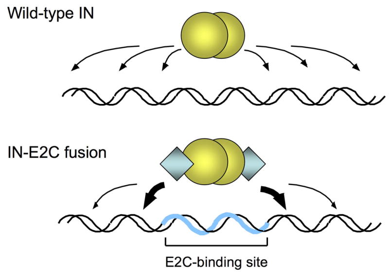
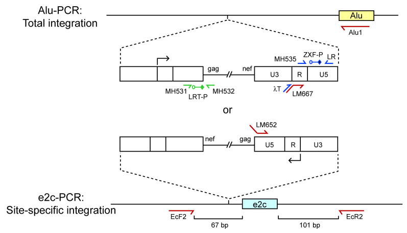
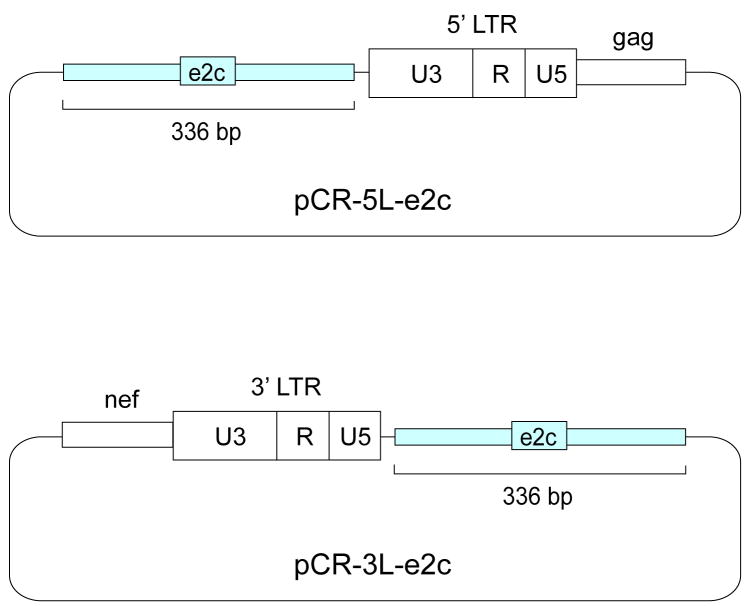
During the life cycle of retroviruses, establishment of a productive infection requires stable joining of a DNA copy of the viral RNA genome into host cell chromosomes. Retroviruses are thus promising vectors for the efficient and stable delivery of genes in therapeutic protocols. Integration of retroviral DNA is catalyzed by the viral enzyme integrase (IN), and one salient feature of retroviral DNA integration is its lack of specificity, as many chromosomal sites can serve as targets for integration. Despite the promise for success in the clinic, one major drawback of the retrovirus-based vector is that any unintended insertion events from the therapy can potentially lead to deleterious effects in patients, as demonstrated by the development of malignancies in both animal and human studies. One approach to directing integration into predetermined DNA sites is fusing IN to a sequence-specific DNA-binding protein, which results in a bias of integration near the recognition site of the fusion partner. Encouraging results have been generated in vitro and in vivo using fusion protein constructs of human immunodeficiency virus type 1 IN and E2C, a designed polydactyl zinc-finger protein that specifically recognizes an 18-base pair DNA sequence. This review focuses on the method for preparing infectious virions containing the IN fusion proteins and on the quantitative PCR assays for determining integration site specificity. Efforts to engineer IN to recognize specific target DNA sequences within the genome may lead to development of effective retroviral vectors that can safely deliver gene-based therapeutics in a clinical setting.


Figures

Fig. 1
Fig. 1
Site-directed integration catalyzed by engineered IN fusion proteins. Integration of retroviral DNA catalyzed by wild-type IN (yellow spheres) is largely non-specific and can occur at various positions along the length of a target DNA (upper panel). In reactions catalyzed by fusion proteins consisting of IN and the designed polydactyl zinc-finger protein E2C (blue diamonds; lower panel), the fusion protein binds specifically to the E2C-recognition sequence e2c (thick blue line) and thereby biases integration in the nearby regions (thick arrows). The e2c site is non-palindromic, and most of the amino acid-DNA contacts are made with the G-rich strand of the target sequence [34, 37]. The IN is depicted as a dimer, but the multimeric state of active IN has not been firmly established.
Fig. 2
Fig. 2
DNA constructs used for trans incorporation of IN-E2C fusion proteins. (A) Fusion protein constructs. Each construct encodes the Vpr protein at the N-terminus (R, open boxes) for packaging of IN (a), IN/E2C (b), or E2C/IN (c) proteins into the viruses. The IN and E2C coding segments are denoted by yellow and blue boxes, respectively. The vertical bars with an open arrowhead denote HIV-1 protease cleavage (PC) sites that are required for removal of Vpr from the IN proteins after packaging [56]. All constructs are encoded in the pLR2P expression plasmid [54]. (B) HIV-1 construct. The plasmid HXB-IN64-Hygro is derived from the HXB2 strain of HIV-1, and contains a defective vpr gene due to an insertion of a T nucleotide at position 5771 [57], and a hygromycin resistance gene (Hygr; red box) in place of env. In addition, IN (yellow box) contains the inactivating Asp to Val substitution at amino acid position 64 (D64V). (C) Envelope expression construct. Each virus was pseudotyped with VSV-G (gray box) driven by the cytomegalovirus (CMV) early promoter [58]. Constructs are not drawn to scale.
Fig. 3
Fig. 3
Trans incorporation of IN-E2C fusion proteins into infectious virions. Producer cells are co-transfected with an HIV expression construct (pHXB-IN64-Hygro) that encodes an IN catalytic mutant (D64V) and hygromycin resistance gene, a fusion protein construct (pLR2P-R-PC-IN or IN-E2C), and an envelope expression construct (pMD.G). After protein expression, the Vpr-fusion protein is packaged into the virus particles via the interaction of Vpr with the p6 portion of Gag.
Fig. 4
Fig. 4
Integration efficiency of viruses containing various IN-E2C fusion proteins. One million HeLa cells were infected for 4 h with equal amounts (ranging from 5–100 ng) of p24 equivalent of the indicated virus (panels A–D). Representative plates of the hygromycin resistance assay are shown. Dark spots on each plate are colonies that grew after selection with 200 μg/ml of hygromycin B for three weeks, beginning two days post-infection. The colonies result from provirus formation and stable expression of the hygromycin resistance gene. The number of resistant colonies per ng p24 for each virus was determined and expressed as a percentage of the colonies produced by the virus containing the WT IN supplied in trans (panel B). The figure is reproduced from previously published data with permission from Copyright © 2006, American Society for Microbiology (doi:10.1128/JVI.80.4.1939-1948.2006).
Fig. 5
Fig. 5
Fluorescence-monitored, real-time nested PCR assays for quantifying integration specificity. HIV-1 cDNA is represented as a thin line flanked by LTRs (open boxes). The arrows above and below the U3-R junction of the LTR indicate the direction of viral transcription. Alu-PCR quantifies proviral DNA integrated in the entire genome, whereas e2c-PCR quantifies proviral DNA integrated near the e2c site on chromosome 17. Proviral integration can be in either orientation and upstream or downstream relative to a specified locus, Alu (yellow box) or e2c (blue box). For simplicity, the diagram depicts only the scenario in which the proviral DNA is integrated in either orientation upstream of an Alu element or the e2c site. Green arrows represent the locations and orientations of the PCR primers for determining the proviral copy number of the Alu-PCR Standard. The quantitative Alu-PCR and e2c-PCR assays consist of two-rounds of PCR. Red arrows represent the locations and orientations of the first-round PCR primers. Alu1 is used for total integration and EcF2 and EcR2 are for e2c-specific integration. Blue arrows represent the locations and orientations of second-round PCR primers, whereas LRT-P (green) and ZXF-P (blue) denote the locations of the fluorescent probes used during real-time PCR. Primers LM652 and LM667 contain phage λ-specific sequences at their 5′ ends, and primer λT anneals specifically to the phage λ sequences.
Fig. 6
Fig. 6
DNA constructs for quantifying proviral DNA integrated upstream or downstream of the e2c site. A 336-bp DNA fragment (blue box) flanking the e2c site, located within the 5′ untranslated region of the erbB-2 gene on human chromosome 17 [49], was amplified by PCR using cellular DNA isolated from uninfected HeLa cells as the template. The PCR product was cloned into the pCR-Blunt II-Topo vector (Invitrogen), resulting in pCR-e2c. An HIV-1 DNA fragment (open box) containing the upstream LTR and part of the gag sequence or the downstream LTR and part of the nef sequence was obtained by PCR. The LTR-gag fragment was cloned into pCR-e2c downstream of the 336-bp e2c-containing fragment, resulting in pCR-5L-e2c. The LTR-nef fragment was cloned upstream of the e2c-containing fragment to form pCR-3L-e2c.

Similar articles

Cited by

References

    1. Langer R. Nature. 1998;392 (6679 Suppl):5–10. - PubMed
    1. Kay MA, Glorioso JC, Naldini L. Nat Med. 2001;7:33–40. - PubMed
    1. Thomas CE, Ehrhardt A, Kay MA. Nat Rev Genet. 2003;4:346–58. - PubMed
    1. Brown PO. In: Retroviruses. Coffin JM, Hughes SH, Varmus HE, editors. Cold Spring Harbor Laboratory Press; Cold Spring Harbor: 1997. pp. 161–203. - PubMed
    1. Kordower JH, Emborg ME, Bloch J, Ma SY, Chu Y, Leventhal L, McBride J, Chen EY, Palfi S, Roitberg BZ, Brown WD, Holden JE, Pyzalski R, Taylor MD, Carvey P, Ling ZD, Trono D, Hantraye P, Deglon N, Aebischer P. Science. 2000;290:767–73. - PubMed

Publication types

LinkOut - more resources