

Diffusion-weighted magnetic resonance imaging as a cancer biomarker: consensus and recommendations
- PMID: 19186405
- PMCID: PMC2631136
- DOI: 10.1593/neo.81328
Diffusion-weighted magnetic resonance imaging as a cancer biomarker: consensus and recommendations
Abstract
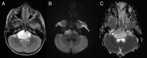
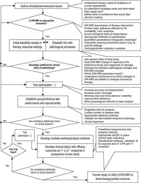
On May 3, 2008, a National Cancer Institute (NCI)-sponsored open consensus conference was held in Toronto, Ontario, Canada, during the 2008 International Society for Magnetic Resonance in Medicine Meeting. Approximately 100 experts and stakeholders summarized the current understanding of diffusion-weighted magnetic resonance imaging (DW-MRI) and reached consensus on the use of DW-MRI as a cancer imaging biomarker. DW-MRI should be tested as an imaging biomarker in the context of well-defined clinical trials, by adding DW-MRI to existing NCI-sponsored trials, particularly those with tissue sampling or survival indicators. Where possible, DW-MRI measurements should be compared with histologic indices including cellularity and tissue response. There is a need for tissue equivalent diffusivity phantoms; meanwhile, simple fluid-filled phantoms should be used. Monoexponential assessments of apparent diffusion coefficient values should use two b values (>100 and between 500 and 1000 mm2/sec depending on the application). Free breathing with multiple acquisitions is superior to complex gating techniques. Baseline patient reproducibility studies should be part of study designs. Both region of interest and histogram analysis of apparent diffusion coefficient measurements should be obtained. Standards for measurement, analysis, and display are needed. Annotated data from validation studies (along with outcome measures) should be made publicly available. Magnetic resonance imaging vendors should be engaged in this process. The NCI should establish a task force of experts (physicists, radiologists, and oncologists) to plan, organize technical aspects, and conduct pilot trials. The American College of Radiology Imaging Network infrastructure may be suitable for these purposes. There is an extraordinary opportunity for DW-MRI to evolve into a clinically valuable imaging tool, potentially important for drug development.
Figures


References
-
- Rudin M. Imaging readouts as biomarkers or surrogate parameters for the assessment of therapeutic interventions. Eur Radiol. 2007;17(10):2441–2457. - PubMed
-
- Johnston KC, Wagner DP, Wang XQ, Newman GC, Thijs V, Sen S, Warach S. Validation of an acute ischemic stroke model: does diffusion-weighted imaging lesion volume offer a clinically significant improvement in prediction of outcome? Stroke. 2007;38(6):1820–1825. - PubMed
-
- Chenevert TL, Brunberg JA, Pipe JG. Anisotropic diffusion in human white matter: demonstration with MR techniques in vivo. Radiology. 1990;177(2):401–405. - PubMed
-
- Stejskal EO, Tanner JE. Spin diffusion measurements: spin echoes in the presence of a time dependent field gradient. J Chem Phys. 1965;42:288–292.
-
- Le Bihan D, Breton E, Lallemand D, Aubin ML, Vignaud J, Laval-Jeantet M. Separation of diffusion and perfusion in intravoxel incoherent motion MR imaging. Radiology. 1988;168:497–505. - PubMed
Publication types
MeSH terms
Substances
LinkOut - more resources
Full Text Sources
Other Literature Sources
Medical