

A direct main olfactory bulb projection to the 'vomeronasal' amygdala in female mice selectively responds to volatile pheromones from males
- PMID: 19187265
- PMCID: PMC2669936
- DOI: 10.1111/j.1460-9568.2009.06638.x
A direct main olfactory bulb projection to the 'vomeronasal' amygdala in female mice selectively responds to volatile pheromones from males
Abstract
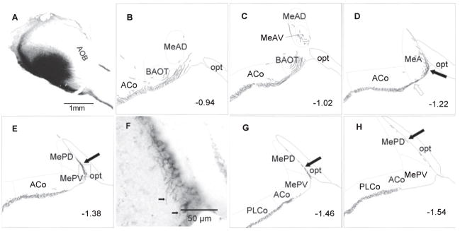
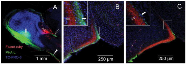
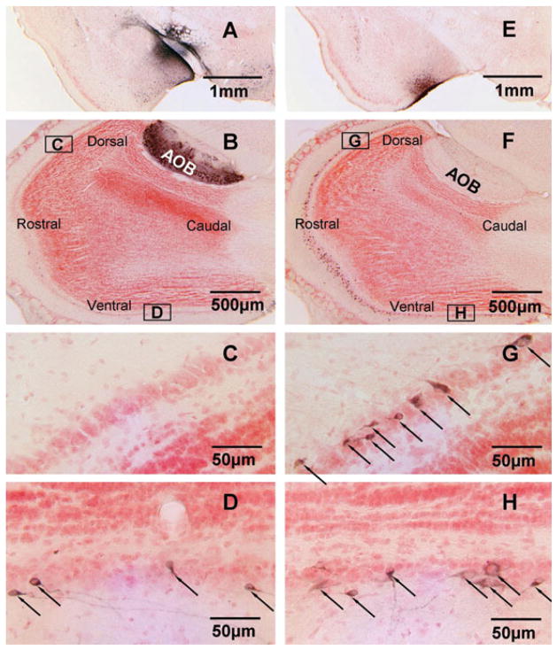
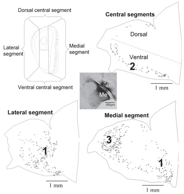
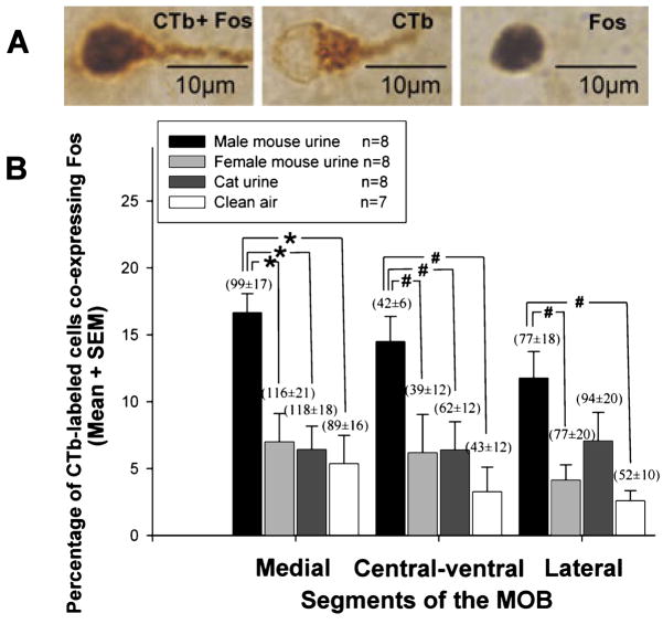
The main olfactory system, like the accessory olfactory system, responds to pheromones involved in social communication. Whereas pheromones detected by the accessory system are transmitted to the hypothalamus via the medial ('vomeronasal') amygdala, the pathway by which pheromones are detected and transmitted by the main system is not well understood. We examined in female mice whether a direct projection from mitral/tufted (M/T) cells in the main olfactory bulb (MOB) to the medial amygdala exists, and whether medial amygdala-projecting M/T cells are activated by volatile urinary odors from conspecifics or a predator (cat). Simultaneous anterograde tracing using Phaseolus vulgaris leucoagglutinin and Fluoro-Ruby placed in the MOB and accessory olfactory bulb (AOB), respectively, revealed that axons of MOB M/T cells projected to superficial laminae of layer Ia in anterior and posterodorsal subdivisions of the medial amygdala, whereas projection neurons from the AOB sent axons to non-overlapping, deeper layer Ia laminae of the same subdivisions. Placement of the retrograde tracer cholera toxin B into the medial amygdala labeled M/T cells that were concentrated in the ventral MOB. Urinary volatiles from male mice, but not from female conspecifics or cat, induced Fos in medial amygdala-projecting MOB M/T cells of female subjects, suggesting that information about male odors is transmitted directly from the MOB to the 'vomeronasal' amygdala. The presence of a direct MOB-to-medial amygdala pathway in mice and other mammals could enable volatile, opposite-sex pheromones to gain privileged access to diencephalic structures that control mate recognition and reproduction.
Figures
Comment in
-
Tracing the scent of a male (Commentary on Kang et al.).Eur J Neurosci. 2009 Feb;29(3):623. doi: 10.1111/j.1460-9568.2009.06667.x. Eur J Neurosci. 2009. PMID: 19222561 No abstract available.
Similar articles
-
A sex comparison of the anatomy and function of the main olfactory bulb-medial amygdala projection in mice.Neuroscience. 2011 Jan 13;172:196-204. doi: 10.1016/j.neuroscience.2010.11.003. Epub 2010 Nov 9. Neuroscience. 2011. PMID: 21070839 Free PMC article.
-
A centrifugal pathway to the mouse accessory olfactory bulb from the medial amygdala conveys gender-specific volatile pheromonal signals.Eur J Neurosci. 2009 Jan;29(2):368-76. doi: 10.1111/j.1460-9568.2008.06564.x. Epub 2008 Dec 11. Eur J Neurosci. 2009. PMID: 19077123 Free PMC article.
-
Sex differences in main olfactory system pathways involved in psychosexual function.Genes Brain Behav. 2020 Feb;19(2):e12618. doi: 10.1111/gbb.12618. Epub 2019 Nov 4. Genes Brain Behav. 2020. PMID: 31634411 Review.
-
Retrograde labelling of mitral/tufted cells in the mouse accessory olfactory bulb following local injections of the lipophilic tracer DiI into the vomeronasal amygdala.Brain Res. 2001 Mar 30;896(1-2):198-203. doi: 10.1016/s0006-8993(01)02225-9. Brain Res. 2001. PMID: 11277993
-
The main and the accessory olfactory systems interact in the control of mate recognition and sexual behavior.Behav Brain Res. 2009 Jun 25;200(2):268-76. doi: 10.1016/j.bbr.2009.01.020. Behav Brain Res. 2009. PMID: 19374011 Review.
Cited by
-
Second-order input to the medial amygdala from olfactory sensory neurons expressing the transduction channel TRPM5.J Comp Neurol. 2012 Jun 1;520(8):1819-30. doi: 10.1002/cne.23015. J Comp Neurol. 2012. PMID: 22120520 Free PMC article.
-
Signal Detection and Coding in the Accessory Olfactory System.Chem Senses. 2018 Nov 1;43(9):667-695. doi: 10.1093/chemse/bjy061. Chem Senses. 2018. PMID: 30256909 Free PMC article. Review.
-
Pheromone-Induced Odor Associative Fear Learning in Rats.Sci Rep. 2018 Dec 7;8(1):17701. doi: 10.1038/s41598-018-36023-w. Sci Rep. 2018. PMID: 30532054 Free PMC article.
-
Human pheromone detection by the vomeronasal organ: unnecessary for mate selection?Chem Senses. 2009 Jul;34(6):529-31. doi: 10.1093/chemse/bjp030. Epub 2009 May 28. Chem Senses. 2009. PMID: 19477954 Free PMC article.
-
Sex-specific processing of social cues in the medial amygdala.Elife. 2014 Jun 3;3:e02743. doi: 10.7554/eLife.02743. Elife. 2014. PMID: 24894465 Free PMC article.
References
-
- Barber PC. Adjacent laminar terminations of two centrifugal afferent pathways to the accessory olfactory bulb in the mouse. Brain Res. 1982;245:215–221. - PubMed
-
- Baxi KN, Dorries KM, Eisthen HL. Is the vomeronasal system really specialized for detecting pheromones? Trends Neurosci. 2006;29:1–7. - PubMed
-
- Beauchamp GK, Martin IG, Wysocki CJ, Wellington JL. Chemoinvestigatory and sexual behavior of male guinea pigs following vomeronasal organ removal. Physiol Behav. 1982;29:329–336. - PubMed
-
- Boehm U, Zou Z, Buck LB. Feedback loops link odor and pheromone signaling with reproduction. Cell. 2005;123:683–695. - PubMed
Publication types
MeSH terms
Substances
Grants and funding
LinkOut - more resources
Full Text Sources
Miscellaneous
