Case Reports
. 2009 Feb 2:7:12.
doi: 10.1186/1477-7819-7-12.

Diagnosis and management of retroperitoneal ancient schwannomas

Affiliations
Case Reports

Diagnosis and management of retroperitoneal ancient schwannomas

Haroon A Choudry et al. World J Surg Oncol. .

Abstract

Background: Ancient schwannomas are degenerate peripheral nerve sheath tumors that very rarely occur in the retroperitoneum. They generally reach large proportions before producing symptoms due to mass effect. We describe three cases of retroperitoneal ancient schwannomas and discuss the diagnosis and management of these tumors.

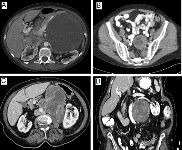
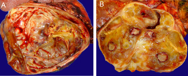
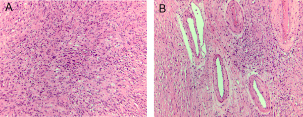
Case presentations: Three female patients with retroperitoneal ancient schwannomas were reviewed. One patient presented with several weeks of upper abdominal pain and lower chest discomfort, whereas back pain and leg pain with associated weakness were predominant symptoms in the remaining two. Abdominal imaging findings demonstrated heterogeneous masses in the retroperitoneum with demarcated margins, concerning for malignancy. The patients successfully had radical excision of their tumors. Histological examination showed encapsulated tumors that displayed alternating areas of dense cellularity and areas of myxoid matrix consistent with a diagnosis of ancient schwannoma.

Conclusion: A diagnosis of ancient schwannoma should be entertained for any heterogeneous, well encapsulated mass in the retroperitoneum. In these cases less radical surgical resection should be considered as malignant transformation of these tumors is extremely rare and recurrence is uncommon following excision.


Figures

Figure 1
Figure 1
Computed tomography findings of three retroperitoneal ancient schwannomas. A. Large partly cystic and solid tumor displacing the pancreas and splenic vein anteriorly and compressing left kidney. Tumor concerning for a sarcoma or pancreatic neoplasm. B. Well defined tumor extending from the retroperitoneum into left pelvis adjacent to sigmoid colon. Region of vascular enhancement around tumor periphery can clearly be seen. C&D. Large heterogeneous enhancing mass adjacent to the aorta and left kidney shown in transverse and sagittal sections. Partial encasement of the aorta and left renal artery is demonstrated concerning for a malignancy.
Figure 2
Figure 2
A. Macroscopic section of large tumor displacing pancreas showing large cystic regions, areas of hemorrhage and calcification B. Tumor showing fibrotic, calcified and cystic regions.
Figure 3
Figure 3
A. The tumor is composed of spindle cell proliferation with hyper-and hypocellular areas and focal cystic degeneration. Rare atypical large nuclei are present in the absence of significant mitotic activity B. Hyalinized and thickened blood vessels can be observed and fibrotic stroma. Immuno-staining was positive for S-100 (not shown).

References

    1. Lane RH, Stephens DH, Reiman HM. Primary retroperitoneal neoplasms: CT findings in 90 cases with clinical and pathologic correlation. AJR Am J Roentgenol. 1989;152:83–89. - PubMed
    1. Loke TK, Yuen NW, Lo KK, Lo J, Chan JC. Retroperitoneal ancient schwannoma: review of clinico-radiological features. Australas Radiol. 1998;42:136–138. doi: 10.1111/j.1440-1673.1998.tb00590.x. - DOI - PubMed
    1. Hide IG, Baudouin CJ, Murray SA, Malcolm AJ. Giant ancient schwannoma of the pelvis. Skeletal Radiol. 2000;29:538–542. doi: 10.1007/s002560000266. - DOI - PubMed
    1. Tortorelli AP, Papa V, Rosa F, Pacelli F, Doglietto GB. Image of the month – retroperitoneal schwannoma. Arch Surg. 2006;141:1259–1261. doi: 10.1001/archsurg.141.12.1259. - DOI - PubMed
    1. Isobe K, Shimizu T, Akahane T, Kato H. Imaging of ancient schwannoma. AJR Am J Roentgenol. 2004;183:331–336. - PubMed

Publication types

LinkOut - more resources