. 2009 Jan 30;33(2):227-36.
doi: 10.1016/j.molcel.2008.12.014.

An unexpected type of ribosomes induced by kasugamycin: a look into ancestral times of protein synthesis?

Affiliations

An unexpected type of ribosomes induced by kasugamycin: a look into ancestral times of protein synthesis?

Anna Chao Kaberdina et al. Mol Cell. .

Abstract

Translation of leaderless mRNAs, lacking ribosomal recruitment signals other than the 5'-terminal AUG-initiating codon, occurs in all three domains of life. Contemporary leaderless mRNAs may therefore be viewed as molecular fossils resembling ancestral mRNAs. Here, we analyzed the phenomenon of sustained translation of a leaderless mRNA in the presence of the antibiotic kasugamycin. Unexpected from the known in vitro effects of the drug, kasugamycin induced the formation of stable approximately 61S ribosomes in vivo, which were proficient in selectively translating leaderless mRNA. 61S particles are devoid of more than six proteins of the small subunit, including the functionally important proteins S1 and S12. The lack of these proteins could be reconciled with structural changes in the 16S rRNA. These studies provide in vivo evidence for the functionality of ribosomes devoid of multiple proteins and shed light on the evolutionary history of ribosomes.


Figures
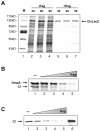
Figure 1
Figure 1. Differential inhibition of lmRNA translation by Ksg in vivo and in vitro
(A) De novo synthesis of a CIΦLacZ fusion protein in the presence of Ksg. Lane 1, protein marker (M). Lanes 2-4, pulse labeling of strain MG1655(pRB381-1) at 30, 60 and 90 min. Lanes 5-7, pulse labeling of strain MG1655(pRB381-1) at 30, 60 and 90 min after addition of Ksg as specified in Experimental Procedures. The molecular masses of marker proteins and the position of the CIΦLacZ fusion protein are indicated on the left and on the right, respectively. (B) Kasugamycin inhibits translation of canonical and lmRNA in vitro. In vitro translation of equimolar amounts of E. coli ompA mRNA and leaderless λ cI mRNA, respectively, with 30S/50S ribosomes and an E. coli S100 extract. Lane 1, translation of both mRNAs in the absence of Ksg. Lane 2-5, translation of both mRNAs in the presence of a 10-fold (lane 2), a 100-fold (lane 3), a 1000-fold (lane 4) and a 10000-fold (lane 5) molar excess of Ksg over ribosomes. The positions of the OmpA and the CI proteins are indicated. (C) Ksg inhibits in vitro translation of λ cI lmRNA by 70S cross-linked ribosomes. Lane 1, translation of cI mRNA in the absence of Ksg; lanes 2-5, translation in the presence of a 10-fold (lane 2), a 100-fold (lane 3), a 1000-fold (lane 4) and a 10000-fold (lane 5) molar excess of Ksg over cross-linked 70S ribosomes. Lane 6, translation of cI mRNA using 30S/50S ribosomes. The position of the CI protein is indicated.
Figure 2
Figure 2. Addition of Ksg leads to the accumulation of protein-deficient 61S and 21S particles in vivo
(A) Ribosome sedimentation profiles of strains MG1655(pRB381) and MG1655(pRB381-1) obtained in the absence of Ksg. (B) and (C) Ribosome sedimentation profiles of the control strain MG1655(pRB381) and MG1655(pRB381-1), respectively, 60 min after addition of 750 μg/ml Ksg. Peaks representing 30S and 50S subunits, 70S ribosomes, and polysomes are indicated. Peaks corresponding to 21S-particles and 61S particles formed in the presence of Ksg are indicated by an open and closed arrow, respectively. The fraction numbers are indicated on the bottom. (D) Protein content of 21S- and 61S particles. The r-proteins were extracted from purified 70S ribosomes, 21S- and 61S particles and identified by quantitative immunoblotting (QI) using antibodies against all r-proteins except S19 as described in Experimental procedures and shown in Figure S2 and in addition subjected to mass spectrometry (MS). The relative abundance of all r-proteins was then normalized with respect to the amount of proteins present in 70S ribosomes. (+) and (−) indicate the presence or absence of r-proteins. (↓) indicates the significant reduction (<50%) of the respective protein. Proteins S15, S17, and S18 were not detected by mass spectrometry.
Figure 3
Figure 3. 61S particles are proficient in translation of lmRNA in vivo
(A) E. coli strain MG1655(pRB381-1) was grown in M9 medium until the OD600 reached 0.2. Then, the culture was divided into two flasks. Sm (100μg/ml) was added to one culture and Ksg (750 μg/ml) to the other. Aliquots of the Sm treated culture were pulse labelled as described in Experimental procedures, 30 (lane 2), 60 (lane 3) and 90 min (lane 4) upon addition of Sm. The Ksg treated culture was incubated for 60 min to allow formation of the 61S particles. Then, Sm (100μg/ml) was added to the culture. 30 min (lane 5), 60 min (lane 6) and 90 min (lane 7) later aliquots of the Ksg/Sm treated culture, respectively, were pulse labelled. Lane 1, pulse labeling of strain MG1655(pRB381-1) at an OD600 of 0.5 in the absence of antibiotics. The position of the CIΦLacZ fusion protein in the autoradiograph is shown. (B) The cI-lacZ lmRNA is present in polysome fractions upon addition of Ksg. Total RNA from polysome fractions (fractions 11-15) of the ribosome profiles shown in Figures 2A and 2C was purified. The presence of leaderless cI-lacZ mRNA and canonical ompA mRNA was determined by primer extension analysis using primers O8 and AvaII (see Table S1), respectively. Lanes 1-5, primer extension signals with total RNA prepared from polysomes of cells grown in the presence of Ksg. Lanes 6-10, extension signals with total RNA prepared from polysomes of cells grown without Ksg. The numbers of the respective fractions (see Figure 2A and C) are given on top of the autoradiograph. Extension signals obtained with in vitro transcribed cI and ompA mRNAs are shown in lane M.
Figure 4
Figure 4. Isolated 61S particles form translation initiation complexes at 5′-terminal start codons and translate lmRNA
Translation initiation complex formation (toeprinting) in the presence of tRNAfMet on leaderless cI mRNA (A) and ompA mRNA (B). (A) Lane 1, primer extension on cI mRNA in the absence of ribosomes and tRNAfMet. Lanes 2, 4 and 6, toeprint analysis with 70S ribosomes, 30S subunits, and 61S particles in the absence of Ksg, respectively. Lanes 3, 5, and 7, toeprint analysis with 70S ribosomes, 30S subunits, and 61S particles in the presence of a 10.000-fold molar excess of Ksg, respectively. The position of the toeprint signal resulting from a translation initiation complex located at position +15 of the cI mRNA is indicated by an arrow. (B) Lane 1, primer extension on ompA mRNA in the absence of ribosomes and tRNAfMet. Lanes 2, 3 and 4, toeprint analysis with 70S ribosomes, 30S subunits, and 61S particles in the absence of Ksg, respectively. The position of the toeprint signal is indicated by an arrow. (C) In vitro translation of cI lmRNA with 61S particles in the absence of Ksg (lane 1) and in the presence of a 100-fold (+, lane 2), and a 10000-fold (++, lane 3) molar excess of Ksg over ribosomes. The position the CI protein is indicated.
Figure 5
Figure 5. Primer extension analyses of the 16S rRNA
(A) The secondary structure of the central portion of the E. coli 16S rRNA, connecting the three major domains of the 30S subunit (head, body, and platform). Helices h2 (red), h24 (green), h26 (cyan), h27 (magenta), and h28 (yellow) which are affected by Ksg in vivo as well as h45 (blue) are highlighted. The packing of the 900 loop of h27 towards the three boxed base-pairs at the bottom of h24 is indicated (Belanger et al., 2004). The binding of primer S1525 (grey line, Table S1) as well as the stop signal obtained in the presence of the modification at A1518/1519 at position 1520 (grey arrowhead) are indicated. The Figure was adapted from the Comparative RNA Web Site (http://www.rna.ccbb.utexas.edu). (B) 16S rRNA purified from 70S ribosomes and 30S subunits isolated from strain MG1655(pRB381-1) grown in the absence of Ksg (Figure 2A, 70S and 30S) as well as 16S rRNA purified from 61S and 21S particles of strain MG1655(pRB381-1) treated with Ksg (Figure 2C, 61S and 21S) was used for primer extension analysis with primer S1525 (Figure 5A; Table S1). The presence of the di-methylation at residues 1518/1519 in the 16S rRNA from 70S ribosomes (lane 1), 30S subunits (lane 2) and 61S particles (lane 3) stopped cDNA synthesis as indicated by the signal (arrow) corresponding to nucleotide 1520 (see also Figure 5A, grey arrowhead). Primer extension of the 16S rRNA from 21S particles did not result in a stop signal, indicating the lack of modification (lane 4). In vitro transcribed 16S rRNA served as an unmodified control (C, lane 5). (C) 70S ribosomes isolated from strain MG1655(pRB381-1) (Figure 2A, 70S) as well as 70S ribosomes and 61S particles from strain MG1655(pRB381-1) treated with Ksg (Figure 2C, 70SK and 61S) were purified as specified in Experimental procedures. Upon DMS treatment the rRNA was isolated and the sites of methylation were determined by primer extension analysis using primer V43, binding to positions 939-955 in 16S rRNA (Table S1). Primer extension analysis of 16S rRNA of 70SK ribosomes (lanes 1 and 2), 61S particles (lanes 3 and 4), and 70S ribosomes (lanes 9 and 10) in the absence of DMS (lanes 1, 3, and 9) or in the presence of DMS (lanes 2, 4, and 10). C, U, A, and G (lanes 5-8): sequencing reactions. The position of helices h2, h27, and h26 are indicated. (D) PhosphorImager quantification of the primer extension analysis shown in (C) of the region spanning h27 (indicated in magenta) and h2 (indicated in red) of 16S rRNA of 70SK ribosomes (red, lane 2), 61S particles (blue, lane 4) and 70S ribosomes (black, lane 10) upon modification by DMS. The corresponding 16S rRNA sequence is shown below.
Figure 6
Figure 6. In vitro formation of 61S particles from 70S ribosomes in the presence of Ksg and lmRNA
Sucrose gradient analysis of 70S ribosomes incubated for 90 min in the absence of cI mRNA and Ksg (black line), and in the presence of either Ksg (cyan line) or cI mRNA (blue line) as specified in Experimental Procedures. The presence of both, Ksg and cI mRNA resulted in formation of 61S particles (red line). A mixture of equimolar amounts of 30S, 50S and 70S served as marker for the position of the subunits and ribosomes (dotted line).
Figure 7
Figure 7. Ksg affects the conformation of the 16S rRNA central pseudoknot
(A) 30S subunit as seen from the subunit interface and enlargement of the region affected by Ksg in vivo. The helices, which are affected by Ksg are highlighted using the same color code as shown in Figure 5A. H26 has been omitted for clarity. Binding of Ksg (sphere model) between the G926 of h28 and the 790 loop of h24 is shown (Schluenzen et al., 2006; Schuwirth et al., 2006). The r-proteins S2 (blue), S6 (purple), S12 (green), S18 (pink), and S21 (yellow) are absent in 61S particles. Nucleoides A1518/A1519 in h45 are shown by blue spheres. (B) 30S subunit shown from the solvent side. H26 (cyan) is located on the platform interacting with proteins S18 (pink) and S1 (not shown). The structures were modeled using PyMOL molecular system software (DeLano, 2002) and PDB file 1VS5 (Schuwirth et al., 2006). (C) Working model for formation of the 61S particle: 70SK ribosomes with a P-site bound fMettRNAfMet (magenta) can form a translation initiation complex on lmRNA (black) containing a 5′-terminal start codon (AUG) but not on a canonical mRNA (blue) containing a Shine and Dalgarno-sequence (SD), as the position of the 5′-UTR of the latter would clash with bound Ksg (yellow sphere; Schluenzen et al., 2006; Schuwirth et al., 2006). At precisely which step Ksg induces the release of ribosomal proteins, i.e. upon formation of the initiation complex or at the beginning of the elongation phase, e.g. upon binding of the charged tRNA cognate to the second codon (blue), remains to be elucidated. At present it is also unknown whether Ksg is released from or remains bound to the 61S particle during translation.

Comment in

Similar articles

Cited by

References

    1. Antoun A, Pavlov MY, Andersson K, Tenson T, Ehrenberg M. The roles of initiation factor 2 and guanosine triphosphate in initiation of protein synthesis. EMBO J. 2003;22:5593–5601. - PMC - PubMed
    1. Backendorf C, Ravensbergen CJ, Van der Plas J, van Boom JH, Veeneman G, Van Duin J. Basepairing potential of the 3′ terminus of 16S RNA: dependence on the functional state of the 30S subunit and the presence of protein S21. Nucleic Acids Res. 1981;9:1425–1444. - PMC - PubMed
    1. Belanger F, Gagnon MG, Steinberg SV, Cunningham PR, Brakier-Gingras L. Study of the functional interaction of the 900 Tetraloop of 16S ribosomal RNA with helix 24 within the bacterial ribosome. J. Mol. Bio.l. 2004;338:683–693. - PubMed
    1. Blattner FR, Plunkett G, 3rd, Bloch CA, Perna NT, Burland V, Riley M, Collado-Vides J, Glasner JD, Rode CK, Mayhew GF, et al. The complete genome sequence of Escherichia coli K-12. Science. 1997;277:1453–1474. - PubMed
    1. Boni IV, Isaeva DM, Musychenko ML, Tzareva NV. Ribosome-messenger recognition: mRNA target sites for ribosomal protein S1. Nucleic Acids Res. 1991;19:155–162. - PMC - PubMed

Publication types

MeSH terms