. 2009 Feb;9(2):140-51.
doi: 10.1016/j.cmet.2008.12.005.

Secondary consequences of beta cell inexcitability: identification and prevention in a murine model of K(ATP)-induced neonatal diabetes mellitus

Affiliations

Secondary consequences of beta cell inexcitability: identification and prevention in a murine model of K(ATP)-induced neonatal diabetes mellitus

Maria Sara Remedi et al. Cell Metab. 2009 Feb.

Abstract

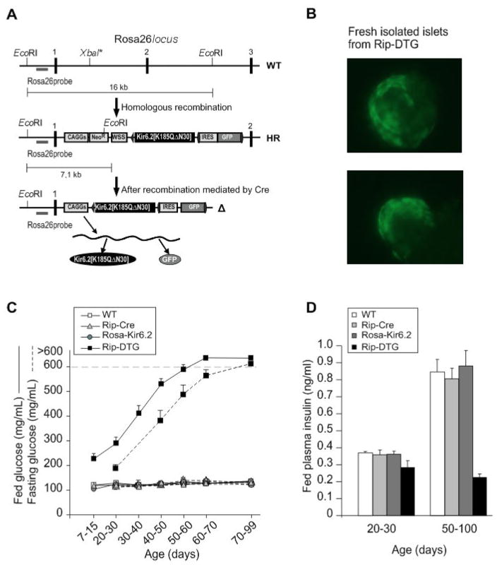
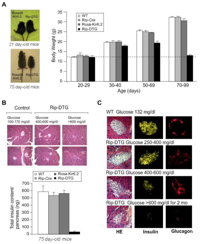
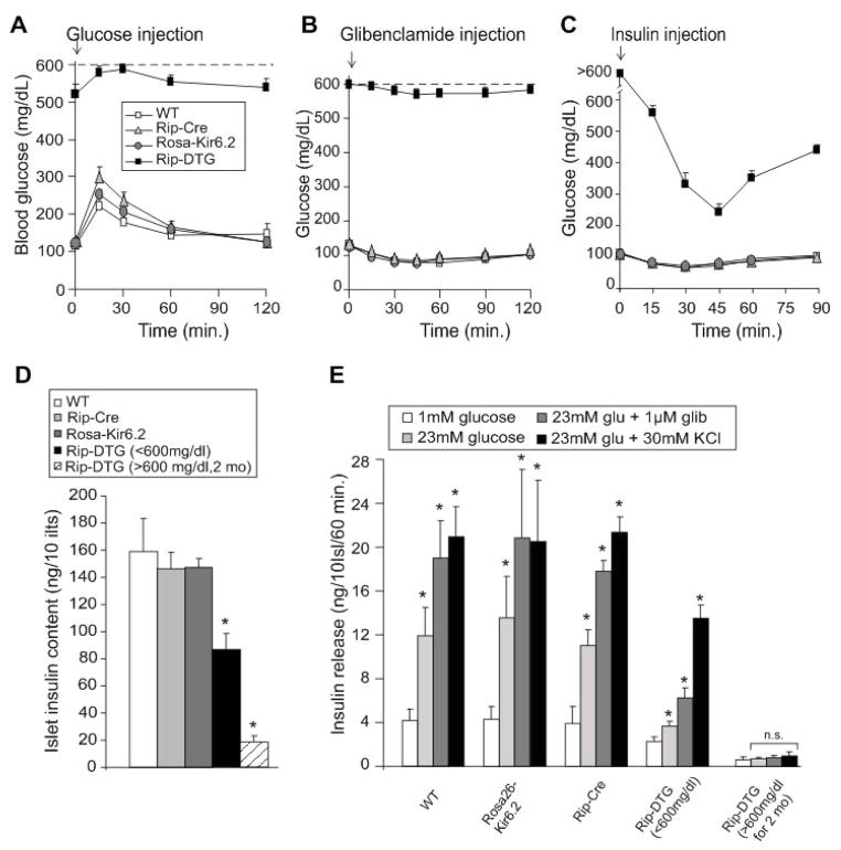
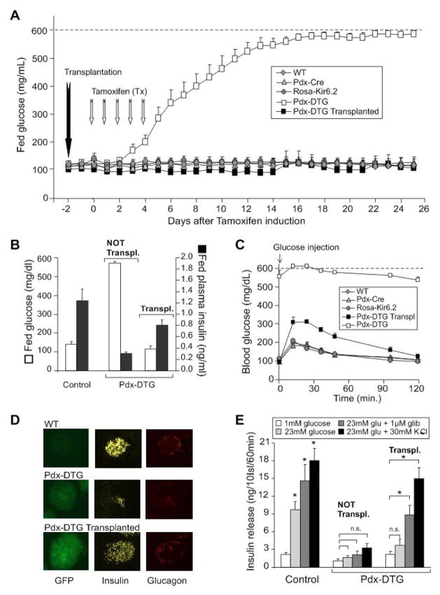
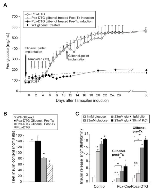
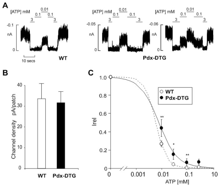
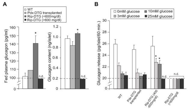
ATP-insensitive K(ATP) channel mutations cause neonatal diabetes mellitus (NDM). To explore the mechanistic etiology, we generated transgenic mice carrying an ATP-insensitive mutant K(ATP) channel subunit. Constitutive expression in pancreatic beta cells caused neonatal hyperglycemia and progression to severe diabetes and growth retardation, with loss of islet insulin content and beta cell architecture. Tamoxifen-induced expression in adult beta cells led to diabetes within 2 weeks, with similar secondary consequences. Diabetes was prevented by transplantation of normal islets under the kidney capsule. Moreover, the endogenous islets maintained normal insulin content and secretion in response to sulfonylureas, but not glucose, consistent with reduced ATP sensitivity of beta cell K(ATP) channels. In NDM, transfer to sulfonylurea therapy is less effective in older patients. This may stem from poor glycemic control or lack of insulin because glibenclamide treatment prior to tamoxifen induction prevented diabetes and secondary complications in mice but failed to halt disease progression after diabetes had developed.


Figures

Figure 1
Figure 1. Induced expression of ATP-insensitive Kir6.2 subunits leads to development of severe diabetes in Rip-DTG mice
(A) Generation of mice with inducible Kir6.2[K185Q,ΔN30] expression. The targeting strategy for generation of a Cre-inducible Kir6.2[K185Q,ΔN30] transgene in the ROSA26 locus is shown. ES cells were transfected with the depicted vector, and homologous recombinants were detected by Southern Blot (not shown). Correctly targeted ES cells were injected into blastocysts. (B) Cre-mediated recombination of the Neo/WSS cassette leads to expression of the Kir6.2[K185Q,ΔN30] protein but also to expression of a GFP via an internal ribosome entry site (IRES). Freshly isolated islets from Rip-Cre/Rosa26-Kir6.2[K185Q,ΔN30] (Rip-DTG) animals show diffuse green fluorescence in the core of the islet, reflecting transgene expression in β-cells. (C) Fed (solid) and fasted (dashed) blood glucose levels from Rip-DTG and control (WT, Rip-Cre and Rosa26-Kir6.2[K185Q,ΔN30] (Rosa-Kir6.2)) littermates over time (n=8–10 mice in each group, mean ± SEM). (D) Fed plasma insulin from all 4 genotype mice at 20–30 day-old and 50–100 day-old.
Figure 2
Figure 2. Growth retardation accompanied by dramatic changes in islet morphology and hormone content in diabetic Rip-DTG animals in late stages of diabetes
(A) (left) Photographs of Rip-DTG mice and single transgenic littermates at 21 days and 75 days. (right). Body weight as a function of age for Rip-DTG, WT and single genotype littermates (n = 12–15 in each case). (B) (top) Low magnification (20x) pancreatic sections from Rip-DTG and control animals stained with hematoxylin-eosin and (lower) pancreatic insulin content as a function of age for Rip-DTG, WT and single genotype littermates (mean ± SEM, n = 4–8 mice in each case). (C) Hematoxylin-eosin staining (left), insulin (middle) and glucagon (right) immunostaining of pancreatic paraffin sections from WT control (top) and Rip-DTG mice at early (middle) and late stages of disease (below).
Figure 3
Figure 3. Insulin sensitivity and secretion in Rip-DTG and control littermate mice
(A) Intraperitoneal glucose tolerance test on fasted WT, Rip-DTG and single transgenic littermate mice at 6 weeks of age. Blood [glucose] versus time following injection of 1.5 g/kg glucose (mean + SEM, n= 8–11 animals in each group). (B, C) Response of fed WT, Rip-DTG and single transgenic littermate mice at 6 weeks of age to single i.p. injection of glibenclamide (1.0 mg/g, B) or response of 6 hours fasted mice to i.p. injection of insulin (0.5 mU/g, C) (mean + SEM, n= 6–10 animals in each group). (D) Insulin content of isolated islets from WT, Rip-DTG and single transgenic littermate mice, at 6 weeks of age, and Rip-DTG also determined at ~3 months of age (glucose >600 mg/dl for > 2 months, dashed bar) (mean ± s.e.m., n= 5–7 animals in each case). (E) Glucose-dependent insulin secretion from isolated islets as in D. Batches of 10 islets were statically pre-incubated with low glucose and then incubated at different glucose concentrations (with or without glibenclamide and K+) for 1hour. Insulin release was measured in the media by radioimmunoassay (n=5–7 mice in each group, samples done in triplicates). Significant differences *p<0.05 between test and WT mice (D) or 1 mM glucose (E) are indicated.
Figure 4
Figure 4. Rapid development of severe diabetes in tamoxifen-induced Pdx-DTG mice, and complete rescue by syngeneic transplant of normal islets
(A) Fed blood glucose levels from Pdx-DTG and control (WT, Pdx-Cre and Rosa-Kir6.2) littermate mice following onset of tamoxifen induction (at 6 weeks of age, n = 6–8 mice in each group). Transplantation of 300 syngeneic islets from single transgene or control littermates was performed in a sub-set of Pdx-DTG animals (n=5, black squares) two days before the onset of tamoxifen induction. (B) Blood glucose and plasma insulin levels in fed Pdx-DTG (transplanted and non-transplanted as indicated) and control littermate mice, 3 weeks following tamoxifen induction (n=5–8 mice in each group). (C) Intraperitoneal glucose tolerance test on Pdx-DTG (transplanted, black squares and non-transplanted, white squares) and control littermate mice. Blood [glucose] versus time following injection of 1.5 g/kg glucose (mean + SEM, n= 4–5 animals in each group). (D) Intrinsic GFP fluorescence of representative freshly isolated islets (left), as well as insulin (middle) and glucagon (right) immunostaining of pancreatic paraffin sections from WT (top) and from transplanted and non-tranplanted Pdx-DTG mice at 3 weeks following induction (below). (E) Glucose-dependent insulin secretion from isolated islets. Batches of 10 islets were statically pre-incubated with low glucose and then incubated at different glucose concentrations (with or without glibenclamide or K+) for 1 hour. Released insulin was measured by radioimmunoassay. n=4–7 mice in each group, samples done in triplicates. Significant differences *p<0.05 between test and 1 mM glucose control are indicated.
Figure 5
Figure 5. Rescue of diabetes and its secondary consequences in Pdx-DTG mice treated with sulfonylureas
(A) Fed blood glucose levels from Pdx-DTG treated with glibenclamide pre- and post-tamoxifen induction respect to controls (WT- glibenclamide treated and Pdx-DTG untreated mice). At 6 weeks of age, Pdx-DTG mice were implanted with slow-release glibenclamide pellets 1-day before (black diamonds) or following onset of tamoxifen induction (grey diamonds) (n = 4 mice in each group). Dashed line shows response of WT mice to glibenclamide implantation (from Remedi and Nichols, 2008) (B) Insulin content of isolated islets from glibenclamide treated WT and Pdx-DTG mice 60 days after tamoxifen induction (mean ± s.e.m., n= 4 animals in each group). (C) Glucose-dependent insulin secretion from isolated islets. Batches of 10 islets were statically pre-incubated with low glucose and then incubated at different glucose concentrations (with or without glibenclamide or K+) for 1 hour. Released insulin was measured by radioimmunoassay. n=3 mice in each group, samples done in triplicates, in Pdx-DTG animals, implanted with glibenclamide pellets (before (pre-) or after (post-) induction, or control littermates. Significant differences *p<0.01 between test and 1 mM glucose control are indicated.
Figure 6
Figure 6. Reduced ATP-sensitivity, but normal density, of KATP currents in primary β-cells from transplanted Pdx-DTG animals
(A) Representative currents recorded from inside-out membrane patches from WT and Pdx-DTG pancreatic β-cells at −50 mV in K-INT solution (see Experimental Procedures). Membrane patches were exposed to different ATP concentrations as shown. (B) KATP channel density and (C) steady-state dependence of membrane current on [ATP] relative to current in zero ATP [Irel]) for KATP channels from wild-type and Pdx-DTG β-cells. Data points represent means + SEM, n= 3 animals (15–30 patches). The fitted lines in C correspond to least squares fits of the Hill equation, with the H (Hill coefficient), and K ½ values allowed to vary. For WT, H= 2, K 1/2= 6 μM (dashed line) and for Pdx-Cre/Rosa-DTG, H= 1.2, and K 1/2= 8 μM (solid line). Significant differences *P < 0.05 and **P < 0.01 from transgenic respect to control cells.
Figure 7
Figure 7. Glucagon content and secretion in Rip-DTG diabetic mice
(A) Plasma glucagon and glucagon content per islet from WT, Rip-DTG and syngeneically transplanted Pdx-DTG mice determined at ~3.5 months of age (mean ± s.e.m., n= 3 animals in each case). Plasma glucagon and glucagon content were determined by radioimmunoassay (see Experimental Procedures). Significant differences *p<0.05 between test and Wt are indicated. (B) Glucose-dependent glucagon secretion from isolated islets as in A. Batches of 10 islets were statically incubated at different glucose concentrations for 1 hour. Glucagon release was measured in the media by radioimmunoassay (n=3 mice in each group, samples done in triplicates). Significant differences *p<0.05 between test and same glucose concentration from controls are indicated.

Similar articles

Cited by

References

    1. Ashcroft FM, Gribble FM. ATP-sensitive K+ channels and insulin secretion: their role in health and disease.[comment] Diabetologia. 1999;42:903–919. - PubMed
    1. Bansal P, Wang Q. Insulin as a physiological modulator of glucagon secretion. Am J Physiol Endocrinol Metab. 2008;295:E751–761. - PubMed
    1. Brown RJ, Sinaii N, Rother KI. Too much glucagon, too little insulin: time course of pancreatic islet dysfunction in new-onset type 1 diabetes. Diabetes Care. 2008;31:1403–1404. - PMC - PubMed
    1. Del Prato S, Marchetti P. Beta- and alpha-cell dysfunction in type 2 diabetes. Horm Metab Res. 2004;36:775–781. - PubMed
    1. Gloyn AL, Cummings EA, Edghill EL, Harries LW, Scott R, Costa T, Temple IK, Hattersley AT, Ellard S. Permanent neonatal diabetes due to paternal germline mosaicism for an activating mutation of the KCNJ11 Gene encoding the Kir6.2 subunit of the beta-cell potassium adenosine triphosphate channel. Journal of Clinical Endocrinology & Metabolism. 2004a;89:3932–3935. - PubMed

Publication types

MeSH terms

LinkOut - more resources