Comparative Study
. 2009 Mar 31;208(1-2):54-60.
doi: 10.1016/j.jneuroim.2009.01.002. Epub 2009 Feb 1.

Intravenous tolerance modulates macrophage classical activation and antigen presentation in experimental autoimmune encephalomyelitis

Affiliations
Comparative Study

Intravenous tolerance modulates macrophage classical activation and antigen presentation in experimental autoimmune encephalomyelitis

Hongmei Li et al. J Neuroimmunol. .

Abstract

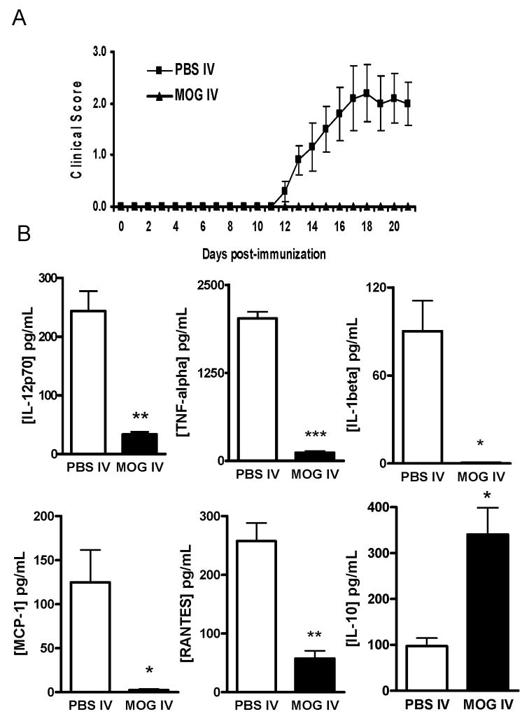
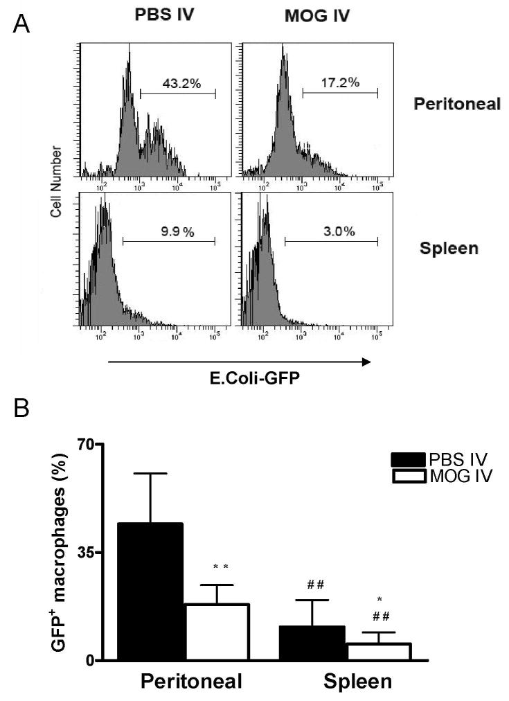
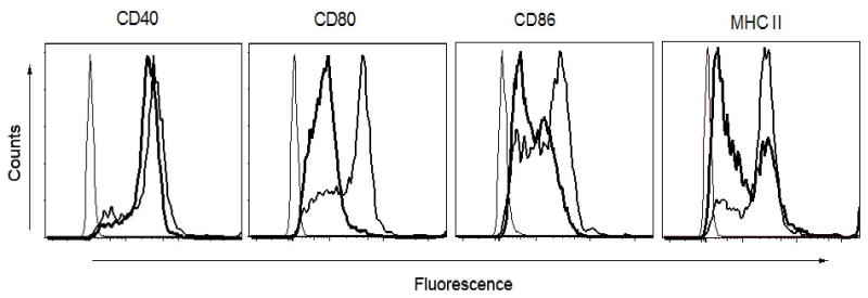
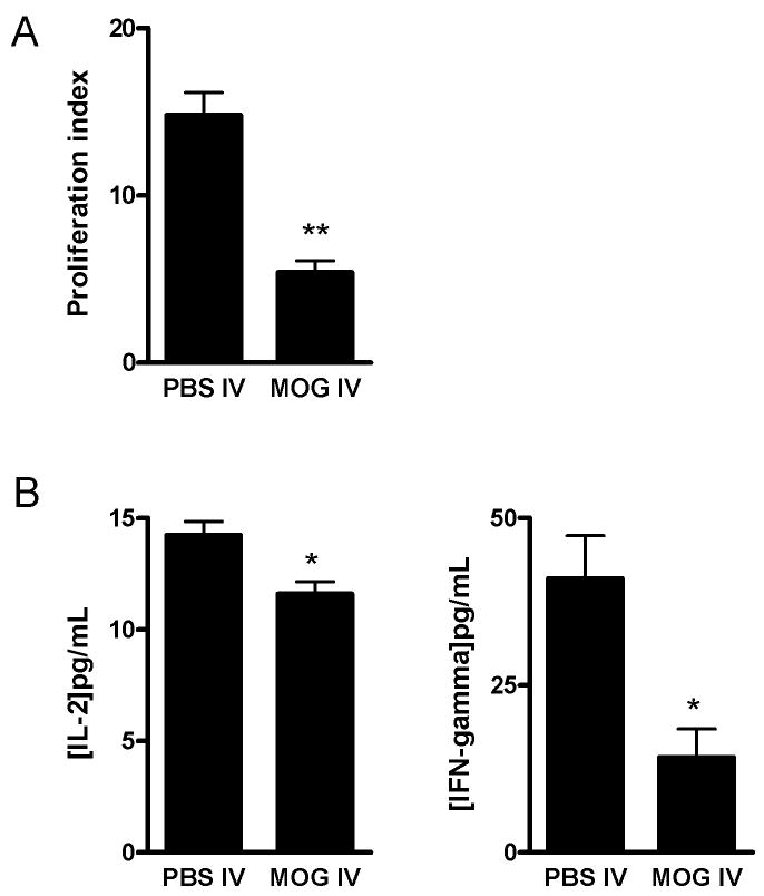
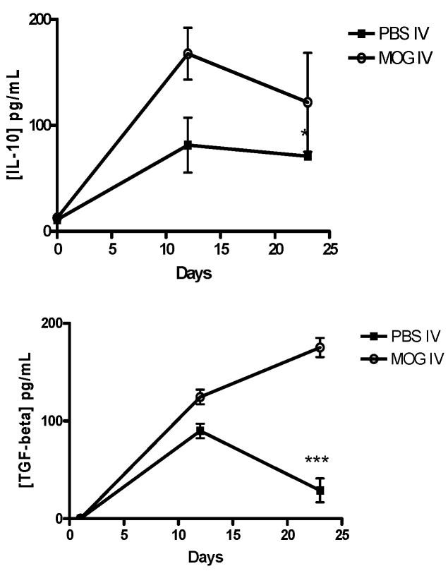
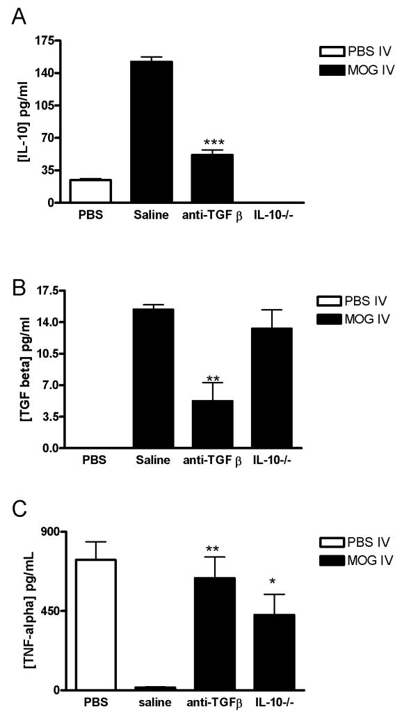
Macrophages act as the first line of self defense by mounting an inflammatory response to antigen and as antigen presenting cells to initiate the adaptive immune response. Inhibition of macrophage activation is one of the possible approaches to modulate inflammation. Intravenous (i.v.) tolerance has proved to be an effective method for ameliorating experimental autoimmune diseases. Whether macrophages are involved in tolerance induction is still largely undefined. In the present study we found that i.v. tolerance induction resulted in lower B7.1, B7.2 and MHC class II molecules, and reduced phagocytosis by both peritoneal macrophages and adherent splenocytes. Macrophages from tolerized mice were associated with a significantly impaired response of MOG-sensitized T cells to MOG. Macrophages from tolerized mice produced low levels of pro-inflammatory molecules IL-12, TNF-alpha, IL-1beta, RANTES and MCP-1 and high levels of IL-10 and TGF-beta. Administration of anti-TGF-beta led to a reduction of IL-10 in tolerized mice. Thus, i.v. tolerance inhibits macrophage classical activation and APC function, increases macrophage alternative activation and IL-10 and TGF-beta production. These cytokines, in turn, induce enhanced production of IL-10 in macrophages in MOG i.v. mice.


Conflict of interest statement

The authors declare no competing financial interest.

Figures

Fig. 1
Fig. 1. Effect of MOG i.v. on cytokine and chemokine release by LPS-stimulated macrophages
(A) I.v. injection of MOG35–55 prevents EAE. C57BL/6 mice were immunized with MOG35–55 + CFA. Pertussis toxin was injected on days 0 and 2 p.i. Two-hundred μg MOG35–55 was injected i.v. on days 3, 5, and 7 p.i. Mice that received i.v. PBS in parallel served as controls. Clinical EAE was scored according to a 0–5 scale (n = 5 in each group). One representative experiment of three is shown (total n = 15 in each group). (B) Peritoneal and splenic macrophages from PBS-i.v. or MOG-i.v. mice were isolated at the end of the experiment as described in Materials and Methods and stimulated with 100 ng/ml LPS for 24 h. The cell supernatants were collected and cytokine and chemokine production was determined by ELISA. Data represent mean ± S.D (n = 5). * p <0.05, ** p <0.01, and *** p <0.001 in comparison between macrophages from MOG-i.v. and PBS-i.v. mice.
Fig. 2
Fig. 2. Phagocytosis of GFP fluorescent transfected E. coli in vitro
Freshly isolated peritoneal macrophages and spleen adherent cells from MOG-i.v. and PBS-i.v. mice were incubated with GFP E. coli for 1hr at 37°C. After washing the cells to remove extracellular E. coli, 1×10⁶ macrophages were analyzed by flow cytometry. (A) GFP-E. coli within the phagocytic cells. Mean values ± S.D (n = 5) were presented in (B). * refers to comparison between PBS-i.v. and MOG-i.v. groups; # refers to comparison between peritoneal macrophages and splenic macrophages in the same groups. *, P<0.05, **, and ##, P<0.01.
Fig. 3
Fig. 3. Effect of MOG i.v. on costimulatory and MHC class II molecule expression in peritoneal macrophages
Macrophages from PBS-i.v. or MOG-i.v. mice were isolated and stimulated with 100 ng/ml LPS for 24 h. The cells were harvested and stained with fluorophore conjugated mAbs against CD40, B7.1, B7.2 and MHC II and analyzed in FACSaria flow cytometer. Bold histogram line represents specific fluorescence for macrophages of MOG-i.v. mice; thinner histogram line represents that of PBS-i.v. EAE mice with isotype-matched control mAbs. One representative experiment of three is shown.
Fig. 4
Fig. 4. Effect of MOG i.v. on the antigen-presenting function of macrophages
Macrophages from PBS-i.v. or MOG-i.v. mice were incubated with LPS for 24 h. These pre-treated macrophages were used as APCs along with T cells as responder cells in the presence of MOG35–55 (25 μg/ml). T cell proliferation was measured after 72 h incubation. Results are presented as proliferation index indicating the absorbance value (at 490 nm) of responder cells (A). IL-2 and IFN-γ production was measured in the culture supernatants after 72 h incubation period (B). Data represent mean ± S.D (n = 9). * p <0.05 and **p < 0.01 in comparison between two groups.
Fig. 5
Fig. 5. Modulated production of TGF-β and IL-10 in ascitic fluids from MOG i.v. mice
The ascitic fluids were collected by peritoneal lavage with 10 ml of saline on indicated days after MOG immunization. Cell-free ascitic fluid was obtained by centrifugation and filtered through a 0.22-pm filter to remove contaminating cells. The cell-free ascitic fluid was stored at −20°C until measurement of cytokine contents. Quantities of TCF-β and IL-10 were measured by ELlSA kits. The results are expressed as the mean with SD of cytokine levels in five individual ascites. Significant at * p < 0.05 or *** p < 0.001 compared with the result between these two groups.
Fig. 6
Fig. 6. Cytokine production from PBS-i.v. and MOG i.v. mice treated with anti-TGF-β
MOG-i.v. C57BL/6 mice were given i.p. anti-TGF-β Ab (50 μg/mouse) from day 12 for 4 consecutive days. Mice that received saline (saline) i.p. in parallel served as controls. MOG-i.v. IL-10−/− mice were injected i.p. with saline. PBS-i.v. mice served as a control. Ascitic fluids were collected as described in Fig. 5 at day 1 after the final anti-TGF-β Ab administration. Production of IL-10 (A) and TGF-β (B) was measured by ELISA. The results are expressed as the mean with SD of cytokine levels in five individual ascites. (C) Peritoneal macrophages were cultured (1 ×10⁶/ml) in the presence or absence of LPS for 24 h in vitro as described in Fig. 1B. Macrophages obtained from PBS-i.v. mice were used as a control. TNF-α concentrations in the supernatant were measured by ELISA. Significant at *p < 0.05, **p < 0.01, ***p <0.001 compared with saline injected MOG-i.v. control mice.

References

    1. Allam R, Anders HJ. The role of innate immunity in autoimmune tissue injury. Curr Opin Rheumatol. 2008;20:538–544. - PubMed
    1. Anderson CF, Gerber JS, Mosser DM. Modulating macrophage function with IgG immune complexes. J Endotoxin Res. 2002;8:477–481. - PubMed
    1. Bhasin JM, Chakrabarti E, Peng DQ, Kulkarni A, Chen X, Smith JD. Sex specific gene regulation and expression QTLs in mouse macrophages from a strain intercross. PLoS ONE. 2008;3:e1435. - PMC - PubMed
    1. Carlsen HS, Yamanaka T, Scott H, Rugtveit J, Brandtzaeg P. The proportion of CD40+ mucosal macrophages is increased in inflammatory bowel disease whereas CD40 ligand (CD154)+ T cells are relatively decreased, suggesting differential modulation of these costimulatory molecules in human gut lamina propria. Inflamm Bowel Dis. 2006;12:1013–1024. - PubMed
    1. Fong CH, Bebien M, Didierlaurent A, Nebauer R, Hussell T, Broide D, Karin M, Lawrence T. An antiinflammatory role for IKKbeta through the inhibition of “classical” macrophage activation. J Exp Med. 2008;205:1269–1276. - PMC - PubMed

Publication types

MeSH terms