. 2009 Feb 1;15(3):933-42.
doi: 10.1158/1078-0432.CCR-08-0399.

MDM2 antagonist nutlin-3 displays antiproliferative and proapoptotic activity in mantle cell lymphoma

Affiliations

MDM2 antagonist nutlin-3 displays antiproliferative and proapoptotic activity in mantle cell lymphoma

Yoko Tabe et al. Clin Cancer Res. .

Abstract

Purpose: Mantle cell lymphoma (MCL) has one of the poorest prognoses of the non-Hodgkin's lymphomas, and novel therapeutic approaches are needed. We wished to determine whether Nutlin-3, a novel small-molecule murine double minute 2 (MDM2) antagonist that efficiently activates TP53, might be effective in inducing cell death in MCL.

Experimental design: MCL cell lines with known TP53 status were treated with Nutlin-3, and biological and biochemical consequences were studied. Synergies with the prototypic genotoxic agent doxorubicin and the novel proteasome inhibitor bortezomib were assessed.

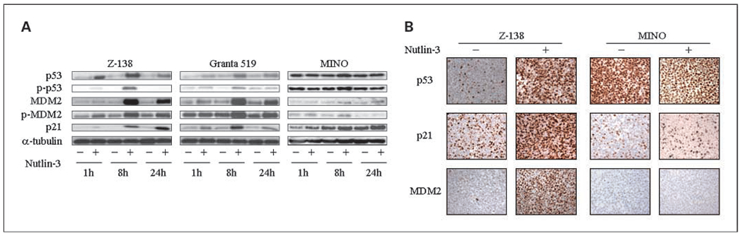
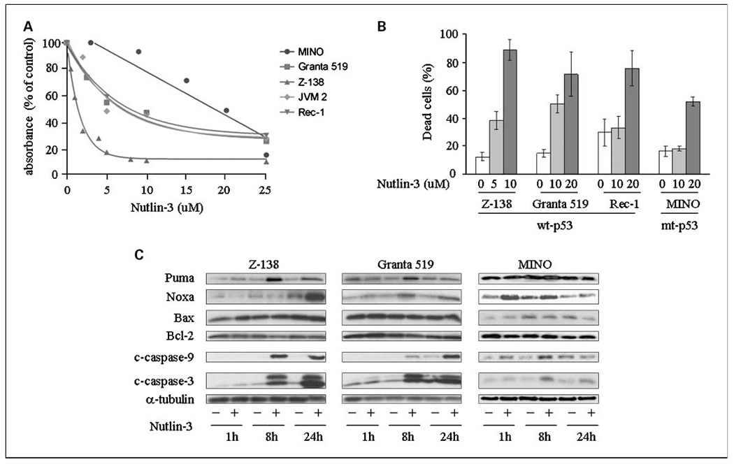
Results: Nutlin-3 resulted in a reduction in cell proliferation/viability (IC50 < 10 micromol/L), an increase in the apoptotic fraction, and cell cycle arrest in wild-type (wt) TP53 Z-138 and Granta 519 cells. These effects were accompanied by TP53 accumulation and induction of TP53-dependent proteins p21, MDM2, Puma, and Noxa. Cell cycle arrest was characterized by suppression of S phase and an increase in the G0-G1 and G2-M fractions and accompanied by suppression of total and phosphorylated retinoblastoma protein and a decrease in G2-M-associated proteins cyclin B and CDC2. The combination of Nutlin-3 with doxorubicin or bortezomib was synergistic in wt-TP53 MCL cells. Nutlin-3 also induced cell cycle arrest and reduced cell viability in the mutant TP53 MINO cells but at a significantly higher IC50 (22.5 micromol/L). These effects were associated with induction of the TP53 homologue p73, slight increases in p21 and Noxa, and caspase activation. Nutlin-3 and bortezomib synergistically inhibited cell growth of MINO.

Conclusion: These findings suggest that the MDM2 antagonist Nutlin-3 may be an effective agent in the treatment of MCL with or without wt-TP53.


Conflict of interest statement

Disclosure of Potential Conflicts of Interest

No potential conflicts of interest were disclosed.

Figures

Fig. 1.
Fig. 1.
Nutlin-3 effects on TP53 and TP53 target proteins. A, Z-138, Granta 519, and MINO cells were treated with Nutlin-3 (Granta 519 and MINO, 10 μmol/L; Z-138, 5 μmol/L). At the indicated time points, cells were lysed and analyzed by Western blot. Carrier (DMSO) alone did not result in any increase in TP53 or TP53 target proteins. The protein levels of TP53 and its traditional targets p21 and MDM2 were increased by Nutlin-3 treatment along with a modest increase in p-TP53 (Ser15) and marked increase in p-MDM2 (Ser166) in Z-138 and Granta 519 cells. A modest increase in p21 is observed in the mt-TP53-bearing cell line MINO. B, immunohistochemistry for TP53, p21, and MDM2 done on paraffin-embedded sections of Z-138 and MINO prepared following treatment with Nutlin-3 (Z-138, 5 μmol/L; MINO, 10 μmol/L) for 8 h. The wt-TP53-bearing Z-138 cell line shows a marked increase in both nuclearTP53 and target proteins p21 and MDM2; no increase in TP53 or in p21 and MDM2 can be discerned by immunohistochemistry in MINO. The high constitutive level of TP53 in MINO is a result of its mutant status.
Fig. 2.
Fig. 2.
Nutlin-3 inhibits cell growth, results in cytotoxicity, and modulates apoptosis-related proteins. A, wt-TP53 MCL cell lines Z-138, Granta 519, Rec-1, and JVM-2 and mt-TP53 MINO cells were exposed to Nutlin-3 at the indicated concentrations for 48 h and cell growth was assessed by the MTS test. Inhibition of cell growth is displayed as percent absorbance of untreated control cells. A significant reduction in absorbance to 50% of control values is seen in the wt-TP53 MCL cell lines by <10 μmol/L Nutlin-3 treatment, whereas the mt-TP53 MINO shows significantly lower sensitivity to Nutlin-3. B, Z-138, Granta 519, Rec-1, and MINO cells were treated with the indicated concentration of Nutlin-3 for 48 h, and the percentage of dead cells was quantified by the trypan blue dye exclusion method. Average + SD of two independent experiments. C, Z-138, Granta 519, and MINO cells were treated with Nutlin-3 (Z-138, 5 μmol/L; Granta 519 and MINO, 10 μmol/L) for the indicated amount of time. Cells were lysed and apoptosis-related proteins were analyzed by Western blot. Nutlin-3 treatment resulted in the induction of both Puma and Noxa, which was closely correlated with caspase activation [accumulation of cleaved caspase (c-caspase) -3 and -9] in wt-TP53 cells. A moderate increase in Noxa and caspase activation is observed in Nutlin-3-treated MINO cells. The other Bcl-2 family proteins, Bax and Bcl-2, remained stable following Nutlin-3 treatment.
Fig. 3.
Fig. 3.
Nutlin-3-induced cell cycle arrest and correlative changes in Rb and Rb phosphorylation. A, percentage of G0-G1, S, and G2-M phases in total viable cells was assessed by cell cycle analysis with PI flow cytometry. S-phase depression was observed at 24 h following treatment with indicated doses of Nutlin-3 in all tested cell lines. B, representative flow cytometric histograms of PI-treated cells after 24 h of Nutlin-3 treatment (Z-138, 1 μmol/L; Granta 519, 10 μmol/L; MINO, 10 μmol/L; BC-1, 5 μmol/L; GM11854, 10 μmol/L). Cell cycle arrest (G0-G1 and/or G2-M) and apoptosis induction (sub-G1) were observed in the tested cell lines. C, Western blot analysis of cell cycle-associated proteins. Z-138, Granta 519, and MINO cells were treated with Nutlin-3 (Z-138, 5 μmol/L; Granta 519 and MINO, 10 μmol/L) for the indicated amount of time. Both phosphorylated (Ser780, Ser795, and Ser807/Ser811) and total Rb levels show a decrease in wt-TP53 Granta 519 and Z-138 but not in mt-TP53 MINO cells. Constitutive expression of cyclin D1 remains unaffected. Cyclin B1 and CDC2 levels are down-regulated between 8 and 24 h following Nutlin-3 treatment in only wt-TP53 cells. D, p73 expression is assessed in Nutlin-3-treated MINO cells by Western blot analysis; p73 level is increased by Nutlin-3 treatment.
Fig. 4.
Fig. 4.
Nutlin-3 treatment does not result in induction of novel TP53 targets PTEN, TSC2, and AMPKβ1. Z-138, Granta 519, and MINO cells were treated with Nutlin-3 (Z-138, 5 μmol/L; Granta 519 and MINO, 10 μmol/L) for the indicated amount of time, and protein expression was analyzed by Western blot. None of the novel TP53 transcriptional targets TSC2, PTEN, or AMPKβ1 showed a significant increase in expression following Nutlin-3 treatment.
Fig. 5.
Fig. 5.
MDMX is not responsible for differential sensitivity to Nutlin-3. Expression level of MDMX was assessed in untreated Z-138, Granta 519, Rec-1, and JVM-2 by Western blot analysis. There was no significant difference of MDMX expression between the highly Nutlin-3-sensitive Z-138 MCL cell line and the other MCL lines.
Fig. 6.
Fig. 6.
Synergistic interaction between Nutlin-3 and bortezomib or doxorubicin in MCL cells. Z-138, Granta 519, and MINO cells were cultured in the presence of escalating doses of Nutlin-3 and bortezomib (A) or Nutlin-3 and doxorubicin (B) using fixed ratios. After 48 h, cell growth inhibition was evaluated by MTS assay and displayed as percent absorbance of untreated control cells.

References

    1. Vousden KH, Lu X. Live or let die: the cell’s response to p53. Nat Rev Cancer 2002;2:594–604. - PubMed
    1. Toledo F, Wahl GM. Regulating the p53 pathway: in vitro hypotheses, in vivo veritas. Nat Rev Cancer 2006;6:909–23. - PubMed
    1. Vassilev LT, Vu BT, Graves B, et al. In vivo activation of the p53 pathway by small-molecule antagonists of MDM2. Science 2004;303:844–8. - PubMed
    1. Petre CE, Sin SH, Dittmer DP. Functional p53 signaling in Kaposi’s sarcoma-associated herpesvirus lymphomas: implications for therapy. J Virol 2007;81:1912–22. - PMC - PubMed
    1. Kojima K, Konopleva M, McQueen T, O’Brien S, Plunkett W, Andreeff M. Mdm2 inhibitor Nutlin-3a induces p53-mediated apoptosis by transcription-dependent and transcription-independent mechanisms and may overcome Atm-mediated resistance to fludarabine in chronic lymphocytic leukemia. Blood 2006;108:993–1000. - PMC - PubMed

MeSH terms