. 2009 Mar 15;587(Pt 6):1331-41.
doi: 10.1113/jphysiol.2008.163311. Epub 2009 Feb 2.

Effect of intracellular lipid droplets on cytosolic Ca2+ and cell death during ischaemia-reperfusion injury in cardiomyocytes

Affiliations

Effect of intracellular lipid droplets on cytosolic Ca2+ and cell death during ischaemia-reperfusion injury in cardiomyocytes

Ignasi Barba et al. J Physiol. .

Abstract

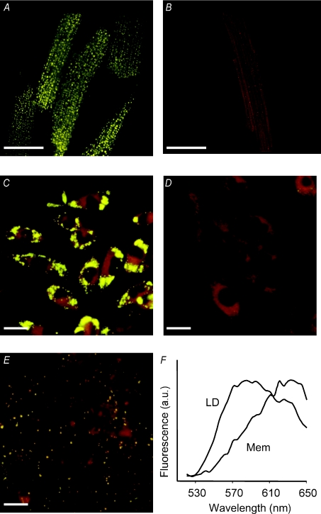
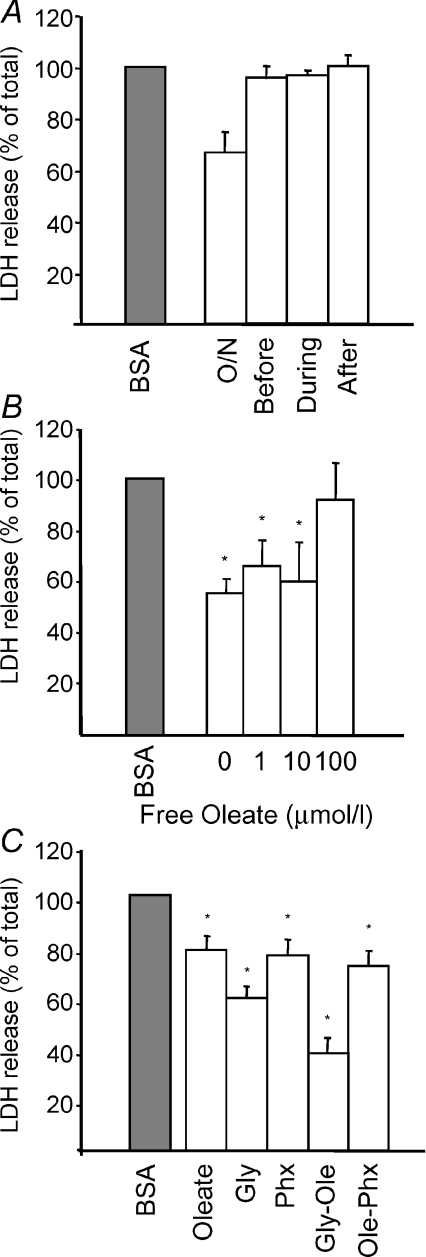
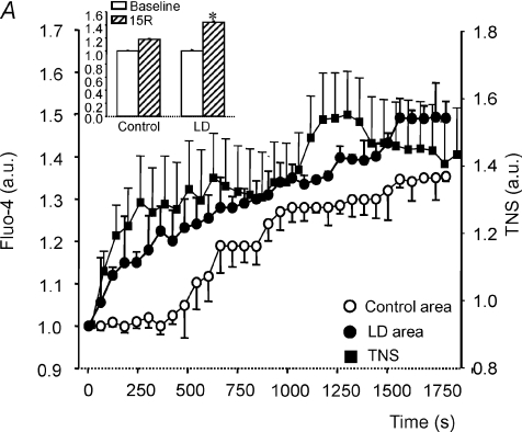
Lipid droplets (LD) consist of accumulations of triacylglycerols and have been proposed to be markers of ischaemic but viable tissue. Previous studies have described the presence of LD in myocardium surviving an acute coronary occlusion. We investigated whether LD may be protective against cell death secondary to ischaemia-reperfusion injury. The addition of oleate-bovine serum albumin complex to freshly isolated adult rat cardiomyocytes or to HL-1 cells resulted in the accumulation of intracellular LD detectable by fluorescence microscopy, flow cytometry and (1)H-nuclear magnetic resonance spectroscopy. Simulated ischaemia-reperfusion of HL-1 cells (respiratory inhibition at pH 6.4 followed by 30 min of reperfusion) resulted in significant cell death (29.7+/-2.6% of total lactate dehydrogenase release). However, cell death was significantly attenuated in cells containing LD (40% reduction in LDH release compared with control cells, P=0.02). The magnitude of LD accumulation was inversely correlated (r(2)=0.68, P=0.0003) with cell death. The protection associated with intracellular LD was not a direct effect of the fatty acids used to induce their formation, because oleate added 30 min before ischaemia, during ischaemia or during reperfusion did not form LD and did not protect against cell death. Increasing the concentration of free oleate during reperfusion progressively decreased the protection afforded by LD. HL-1 cells labelled with fluo-4, a Ca(2+)-sensitive fluorochrome, fluorescence within LD areas increased more throughout simulated ischaemia and reperfusion than in the cytosolic LD-free areas of the same cells. As a consequence, cells with LD showed less cytosolic Ca(2+) overload than control cells. These results suggest that LD exert a protective effect during ischaemia-reperfusion by sequestering free fatty acids and Ca(2+).


Figures

Figure 1
Figure 1. Fluorescent imaging of LD loaded cells
Fluorescence micrographs of adult rat cardiomyocytes supplemented with oleate–BSA (2 mmol l−1; A) or BSA alone (B). Micrographs C, D and E correspond to HL-1 cardiomyocytes supplemented with oleate–BSA, BSA alone and after an overnight hypoxic incubation in 1% O2 without oleate–BSA, respectively. Cells were stained with Nile red at a final concentration of 1 μmol l−1. Lipid droplets appear as bright yellow dots. Scale bars represent 25 μm in A–C and 20 μm in D and E. F shows the emission spectra recorded from areas corresponding to lipid droplets (LD) or membranes (Mem) in HL-1 cells loaded with oleate–BSA.
Figure 2
Figure 2. Flow citometry and H-NMR of LD loaded cells
Flow cytometry dot plots of control HL-1 cardiomyocytes (A) and HL-1 cardiomyocytes treated overnight with 0.2 mmol l−1 oleate–BSA (B). Spectra in C and D correspond to control HL-1 cells and HL-1 cells loaded with 0.2 mmol l−1 oleate–BSA, respectively, and were acquired with a NOESY presat one-dimenstional sequence. Vertical scale in C is increased twofold. E shows the increase in intracellular lipids in response to varying overnight loading concentrations of oleate–BSA measured by flow cytometry (▪) and lipid-to-creatine ratio (◊) in the NMR spectra. F shows an NMR spectrum of an oleate standard acquired with the same sequence as C and D, in which ‘a’ indicates protons from methyl groups of fatty acyl chains –CH3, ‘b’ methylene protons –CH2–, ‘c’ methylene protons close to the ester bond O=C–CH2–CH2–, ‘d’ methylene groups close to a double bond –CH2–CH= and ‘e’ methylene next to the ester bond O=C–CH2–.
Figure 3
Figure 3. Effect of LD on ischemia–reperfusion induced cell death
A, bar graph showing LDH release during normoxia (Nx), simulated ischaemia (SI) and reperfusion (R) in adult rat cardiomyocytes loaded with BSA (open bars) or oleate–BSA (grey bars). B, cellular death during reperfusion in HL-1 cardiomyocytes after different overnight oleate loading concentrations. Results are expressed as the percentage of cell death compared with control cultures. *p < 0.05 compared to BSA loaded cells.
Figure 4
Figure 4. Effect of different interventions on reperfusion induced cell death
A, LDH release in HL-1 cells after simulated ischaemia–reperfusion when 0.2 mmol l−1 oleate–BSA was added 30 min before ischaemia (before), during ischaemia (during) or during reperfusion (after) compared with control BSA-loaded cells (grey bar) and cells loaded overnight with oleate–BSA (O/N). B, cell death at increasing concentrations of free oleate added during reperfusion in cells when lipid droplets had been induced by overnight incubation with oleate–BSA. C, cell death during reperfusion, comparing the effects of LD (overnight oleate–BSA loading), 5 mmol l−1 glycine to inhibit mitochondria transition pore opening, LD and glycine and cells with LD and perhexiline to inhibit the use of fatty acids as energy source. *P < 0.05 compared with control BSA-loaded cells (grey bar).
Figure 5
Figure 5. Effect of LD on Ca2+ overload during ischemia–reperfusion
A, increase in Ca2+ concentration in HL-1 cardiomyocytes submitted to 30 min of simulated ischaemia and 15 min of reperfusion, measured by changes in fluo-4 fluorescence in areas of LD (•), previously defined by light microscopy, versus cytosolic areas devoid of LD (○) within the same cells; the inset represents the relative Ca2+ increase at the end of reperfusion (15R) in those areas. Filled squares indicate whole-cell TNS fluorescence. B, cytosolic area of control cells versus cytosolic area of cells containing LD throughout simulated ischaemia (SI) and reperfusion (R) periods. Results are expressed as means ±s.e.m. of n= 10–14 cells in each group.

References

    1. Awonusonu F, Srinivasan S, Strange J, Al-Jumaily W, Bruce MC. Developmental shift in the relative percentages of fibroblast subsets: role of apoptosis postseptation. Am J Physiol Lung Cell Mol Physiol. 1999;277:L848–L859. - PubMed
    1. Barba I, Cabanas ME, Arus C. The relationship between nuclear magnetic resonance-visible lipids, lipid droplets, and cell proliferation in cultured C6 cells. Cancer Res. 1999;59:1861–1868. - PubMed
    1. Barrabes JA, Garcia-Dorado D, Ruiz-Meana M, Piper HM, Solares J, Gonzalez MA, Oliveras J, Herrejon MP, Soler J. Myocardial segment shrinkage during coronary reperfusion in situ. Relation to hypercontracture and myocardial necrosis. Pflugers Arch. 1996;431:519–526. - PubMed
    1. Bianchi K, Rimessi A, Prandini A, Szabadkai G, Rizzuto R. Calcium and mitochondria: mechanisms and functions of a troubled relationship. Biochim Biophys Acta. 2004;1742:119–131. - PubMed
    1. Bush KT, Lee H, Nagele RG. Lipid droplets of neuroepithelial cells are a major calcium storage site during neural tube formation in chick and mouse embryos. Experientia. 1992;48:516–519. - PubMed

Publication types

MeSH terms