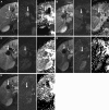
Unresectable hepatocellular carcinoma: serial early vascular and cellular changes after transarterial chemoembolization as detected with MR imaging
- PMID: 19188315
- PMCID: PMC6944072
- DOI: 10.1148/radiol.2502072222
Unresectable hepatocellular carcinoma: serial early vascular and cellular changes after transarterial chemoembolization as detected with MR imaging
Abstract
Purpose: To prospectively assess serial changes in contrast material-enhanced and diffusion-weighted (DW) magnetic resonance (MR) imaging values within 1 month after transarterial chemoembolization (TACE) in patients with unresectable hepatocellular carcinoma (HCC).
Materials and methods: Institutional review board approval was obtained for this prospective HIPAA-compliant study. MR imaging was performed before and within 24 hours after TACE in 24 patients with HCC (21 male, three female; mean age, 59 years and 62 years, respectively). Serial MR imaging was subsequently performed 1, 2, 3, and 4 weeks after therapy. The imaging protocol included fast spin-echo T2-weighted MR imaging, breath-hold DW echo-planar MR imaging, and breath-hold unenhanced and contrast-enhanced T1-weighted three-dimensional fat-suppressed gradient-recalled-echo MR imaging in the arterial and portal venous phases. Tumor size, enhancement, and apparent diffusion coefficient (ADC) values were recorded before and sequentially after treatment. Regression models for the correlated data were used to assess changes in these parameters over time after TACE.
Results: Mean tumor size was 7.5 cm and was unchanged up to 4 weeks after therapy. Reduction in tumor enhancement in the arterial phase occurred immediately after TACE, with a consistent reduction occurring 1-3 weeks after therapy (P = .001). Reduction in tumor enhancement in the portal venous phase also occurred immediately after TACE, with a consistent reduction occurring 1-3 weeks after therapy (P = .0003). The increase in tumor ADC value was significant 1-2 weeks after therapy (P = .004), borderline significant 3 weeks after therapy, and insignificant 24 hours and 4 weeks after therapy.
Conclusion: Significant reduction in tumor enhancement occurred within 24 hours after TACE and persisted up to 4 weeks after TACE. Lesser changes in the ADC value appeared 1 week after TACE, persisted through 2 weeks after TACE, and became less apparent 3 and 4 weeks after TACE. No change in tumor size was recorded during the follow-up period.
Figures


Comment in
-
Unresectable hepatocellular carcinoma: serial early vascular and cellular changes after transarterial chemoembolization.Radiology. 2009 Feb;250(2):324-6. doi: 10.1148/radiol.2502081988. Radiology. 2009. PMID: 19188308 No abstract available.
Similar articles
-
The role of functional MR imaging in the assessment of tumor response after chemoembolization in patients with hepatocellular carcinoma.J Vasc Interv Radiol. 2006 Mar;17(3):505-12. doi: 10.1097/01.RVI.0000200052.02183.92. J Vasc Interv Radiol. 2006. PMID: 16567675
-
Radiologic-pathologic analysis of contrast-enhanced and diffusion-weighted MR imaging in patients with HCC after TACE: diagnostic accuracy of 3D quantitative image analysis.Radiology. 2014 Dec;273(3):746-58. doi: 10.1148/radiol.14140033. Epub 2014 Jul 15. Radiology. 2014. PMID: 25028783 Free PMC article.
-
Role of functional magnetic resonance imaging in assessing metastatic leiomyosarcoma response to chemoembolization.J Comput Assist Tomogr. 2008 May-Jun;32(3):347-52. doi: 10.1097/RCT.0b013e318134ecd6. J Comput Assist Tomogr. 2008. PMID: 18520535
-
A Review Of Mri Appearances Of Lipiodol In Conventional Tace (Ctace) Treated Hepatocellular Carcinomas.J Ayub Med Coll Abbottabad. 2023 Oct-Dec;35(Suppl 1)(4):S740-S745. doi: 10.55519/JAMC-S4-11966. J Ayub Med Coll Abbottabad. 2023. PMID: 38406903 Review.
-
Contrast-enhanced Ultrasound Identifies Patent Feeding Vessels in Transarterial Chemoembolization Patients With Residual Tumor Vascularity.Ultrasound Q. 2020 Sep;36(3):218-223. doi: 10.1097/RUQ.0000000000000513. Ultrasound Q. 2020. PMID: 32890324 Free PMC article. Review.
Cited by
-
Can C-arm cone-beam CT detect a micro-embolic effect after TheraSphere radioembolization of neuroendocrine and carcinoid liver metastasis?Cancer Biother Radiopharm. 2013 Jul-Aug;28(6):459-65. doi: 10.1089/cbr.2012.1390. Cancer Biother Radiopharm. 2013. PMID: 23484809 Free PMC article. Clinical Trial.
-
Intravoxel incoherent motion model-based analysis of diffusion-weighted magnetic resonance imaging with 3 b-values for response assessment in locoregional therapy of hepatocellular carcinoma.Onco Targets Ther. 2016 Oct 19;9:6425-6433. doi: 10.2147/OTT.S113909. eCollection 2016. Onco Targets Ther. 2016. PMID: 27799790 Free PMC article.
-
Prognostic value of baseline volumetric multiparametric MR imaging in neuroendocrine liver metastases treated with transarterial chemoembolization.Eur Radiol. 2019 Oct;29(10):5160-5171. doi: 10.1007/s00330-019-06100-3. Epub 2019 Mar 15. Eur Radiol. 2019. PMID: 30877462
-
Diffusion-weighted imaging of the liver: techniques and applications.Magn Reson Imaging Clin N Am. 2014 Aug;22(3):373-95. doi: 10.1016/j.mric.2014.04.009. Magn Reson Imaging Clin N Am. 2014. PMID: 25086935 Free PMC article. Review.
-
Three-dimensional Image-based Mechanical Modeling for Predicting the Response of Breast Cancer to Neoadjuvant Therapy.Comput Methods Appl Mech Eng. 2017 Feb 1;314:494-512. doi: 10.1016/j.cma.2016.08.024. Epub 2016 Sep 1. Comput Methods Appl Mech Eng. 2017. PMID: 28042181 Free PMC article.
References
-
- El-Serag HB, Mason AC. Rising incidence of hepatocellular carcinoma in the United States. N Engl J Med 1999;340:745–750. - PubMed
-
- American Cancer Society. Cancer facts and figures. Atlanta, Ga: American Cancer Society, 2006.
-
- Carr BI Hepatic artery chemoembolization for advanced stage HCC: experience of 650 patients. Hepatogastroenterology 2002;49:79–86. - PubMed
-
- Ahmed M, Goldberg SN. Thermal ablation therapy for hepatocellular carcinoma. J Vasc Interv Radiol 2002;13(9 pt 2):S231–S244. - PubMed
-
- Ramsey DE, Kernagis LY, Soulen MC, Geschwind JF. Chemoembolization of hepatocellular carcinoma. J Vasc Interv Radiol 2002;13(9 pt 2):S211–S221. - PubMed
Publication types
MeSH terms
Substances
Grants and funding
LinkOut - more resources
Full Text Sources
Other Literature Sources
Medical
Miscellaneous

