. 2009 Apr 10;284(15):9788-95.
doi: 10.1074/jbc.M809339200. Epub 2009 Feb 2.

Two dermatan sulfate epimerases form iduronic acid domains in dermatan sulfate

Affiliations

Two dermatan sulfate epimerases form iduronic acid domains in dermatan sulfate

Benny Pacheco et al. J Biol Chem. .

Abstract

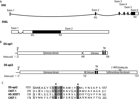
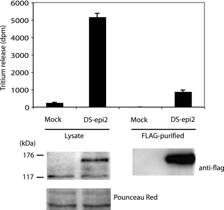
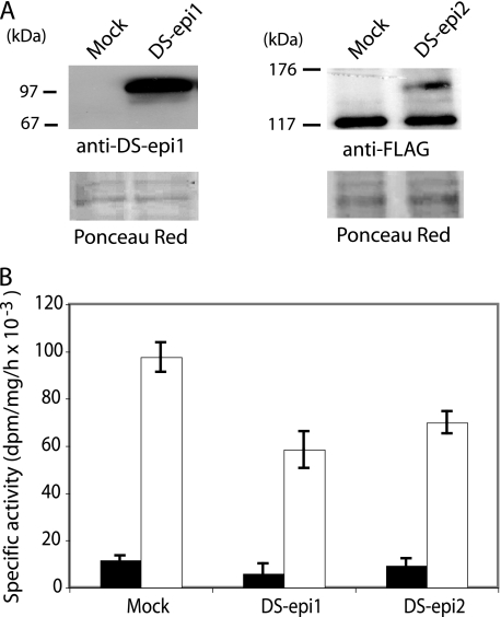
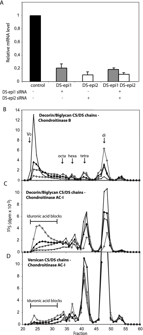
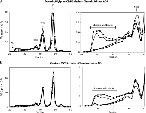
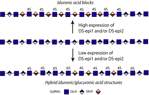
A second dermatan sulfate epimerase (DS-epi2) was identified as a homolog of the first epimerase (DS-epi1), which was previously described by our group. DS-epi2 is 1,222 amino acids long and has an approximately 700-amino acid N-terminal epimerase domain that is highly conserved between the two enzymes. In addition, the C-terminal portion is predicted to be an O-sulfotransferase domain. In this study we found that DS-epi2 has epimerase activity, which involves conversion of d-glucuronic acid to l-iduronic acid (EC 5.1.3.19), but no O-sulfotransferase activity was detected. In dermatan sulfate, iduronic acid residues are either clustered together in blocks or alternating with glucuronic acid, forming hybrid structures. By using a short interfering RNA approach, we found that DS-epi2 and DS-epi1 are both involved in the biosynthesis of the iduronic acid blocks in fibroblasts and that DS-epi2 can also synthesize the hybrid structures. Both iduronic acid-containing domains have been shown to bind to several growth factors, many of which have biological roles in brain development. DS-epi2 has been genetically linked to bipolar disorder, which suggests that the dermatan sulfate domains generated by a defective enzyme may be involved in the etiology of the disease.


Figures

FIGURE 1.
FIGURE 1.
Gene and domain structure of the DS epimerases. A, schematic view of the DSE and DSEL genomic structure. Exons are shown with rectangles, and introns with curved lines. Coding regions are shown as filled boxes. B, domain structure of the two epimerases, showing a common N-terminal epimerase domain followed by two distinct domains of different length (of unknown function) and, in the case of DS-epi2, by an O-sulfotransferase-like domain. The locations of signal peptides (SP) and predicted transmembrane regions (TM) are also shown. A potential 3′-PAPS-binding site is depicted with a straight line. C, amino acid conservation of the 3′-PAPS-binding site is shown by alignment (generated with ClustalW) to the protein sequences of human C6ST-1, HS-3OST1, and C4ST-1. Catalytic amino acids are marked with asterisks.
FIGURE 2.
FIGURE 2.
DS-epi2 has epimerase activity. Epithelial 293HEK cells were transiently transfected with empty pCS2 expression vector or with the vector containing DS-epi2 in-frame with an N-terminal FLAG tag. Top, after 48 h, cell lysates were prepared, and 280 μg of desalted proteins was assayed. Alternatively, 2 mg of lysate was immunopurified by an anti-FLAG M2-agarose gel, and one-fifth of the resuspended gel-bound material was used in each assay. Data represent mean ± 2 S.D. of triplicates. Repeated transfections (n = 12) ranged from 12- to 35-fold overexpression of epimerase activity, compared with mock controls. B, 20 μg of lysates or one-fifth of the immunopurified gel-bound material was subjected to Western blot and probed with peroxidase-conjugated anti-FLAG antibody.
FIGURE 3.
FIGURE 3.
Analysis for O-sulfotransferase activity of DS-epi2. Microsomes were prepared from 293HEK cells overexpressing full-length DS-epi1 or DS-epi2 and resuspended in dialysis buffer without detergent. A, Western blot of 25 μg of microsomal preparations. B, O-sulfotransferase activity was assayed (mean ± 2 S.D. of triplicates) without detergent (black bars) or with detergent (empty bars). The O-sulfotransferase assay shown was performed at pH 6.8 using chondroitin as substrate. Twenty-five μg of microsomes from mock cells incorporated 2,700 dpm, when assayed with detergent.
FIGURE 4.
FIGURE 4.
Endogenous DS-epi1 and DS-epi2 are both active in the biosynthesis of iduronic acid blocks, as revealed by siRNA-mediated down-regulation in human lung fibroblasts. DS-epi1 and DS-epi2 were down-regulated by siRNA treatment, either individually or in combination. A, relative qRT-PCR quantification of DS-epi1 mRNA (grey bars) or DS-epi2 mRNA (empty bars) extracted from the cells after the labeling period. Data represent expression relative to that in the control cells and show mean ± 2 S.D. of triplicates. B–D, labeled proteoglycans were size-fractionated into versican and a mixture of decorin and biglycan. Purified CS/DS chains were cleaved with chondroitinase B or chondroitinase AC-I, as indicated. Split products were separated on the size-permeation Superdex Peptide column. Elution positions of di-, tetra-, hexa-, and octasaccharides are indicated by arrows. AC-I-resistant iduronic acid blocks are shown. Diamonds, control; squares, DS-epi1 siRNA; triangles, DS-epi2 siRNA; circles, DS-epi1 and DS-epi2 siRNAs.
FIGURE 5.
FIGURE 5.
Differences in epimerization pattern obtained after overexpression of DS-epi1 or DS-epi2 in epithelial 293HEK cells. Full-length DS-epi1 or DS-epi2 was overexpressed in 293HEK cells. Labeled CS/DS chains were obtained from purified versican or decorin/biglycan proteoglycans. Purified CS/DS chains were cleaved with chondroitinase AC-I, and split products were separated as in Fig. 4. On the left, the full chromatograms are shown, and on the right a 6× zoom of the fractions equal or larger than hexasaccharides is presented. Diamonds, control; squares, DS-epi1; triangles, DS-epi2; circles, DS-epi1 and DS-epi2.
FIGURE 6.
FIGURE 6.
Formation of different iduronic acid domains by DS-epi1 and DS-epi2.

References

    1. Sugahara, K., and Mikami, T. (2007) Curr. Opin. Struct. Biol. 17 536–545 - PubMed
    1. Trowbridge, J. M., and Gallo, R. L. (2002) Glycobiology 12 117R–125R - PubMed
    1. Prabhakar, V., and Sasisekharan, R. (2006) Adv. Pharmacol. 53 69–115 - PubMed
    1. Malmstrom, A. (1984) J. Biol. Chem. 259 161–165 - PubMed
    1. Maccarana, M., Olander, B., Malmstrom, J., Tiedemann, K., Aebersold, R., Lindahl, U., Li, J. P., and Malmstrom, A. (2006) J. Biol. Chem. 281 11560–11568 - PubMed

Publication types

LinkOut - more resources