

Kinetics of GLUT4 trafficking in rat and human skeletal muscle
- PMID: 19188436
- PMCID: PMC2661600
- DOI: 10.2337/db08-1539
Kinetics of GLUT4 trafficking in rat and human skeletal muscle
Abstract
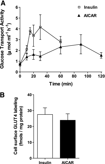
Objective: In skeletal muscle, insulin stimulates glucose transport activity three- to fourfold, and a large part of this stimulation is associated with a net translocation of GLUT4 from an intracellular compartment to the cell surface. We examined the extent to which insulin or the AMP-activated protein kinase activator AICAR can lead to a stimulation of the exocytosis limb of the GLUT4 translocation pathway and thereby account for the net increase in glucose transport activity.
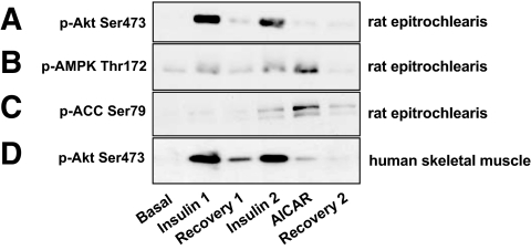
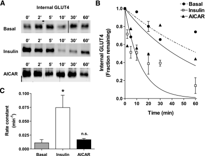
Research design and methods: Using a biotinylated photoaffinity label, we tagged endogenous GLUT4 and studied the kinetics of exocytosis of the tagged protein in rat and human skeletal muscle in response to insulin or AICAR. Isolated epitrochlearis muscles were obtained from male Wistar rats. Vastus lateralis skeletal muscle strips were prepared from open muscle biopsies obtained from six healthy men (age 39 +/- 11 years and BMI 25.8 +/- 0.8 kg/m2).
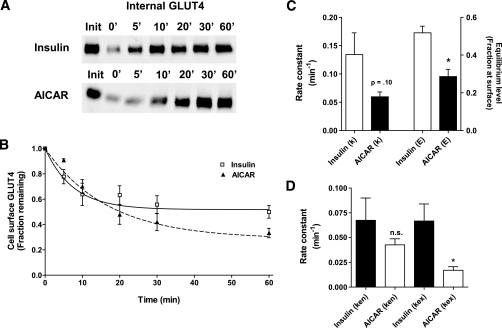
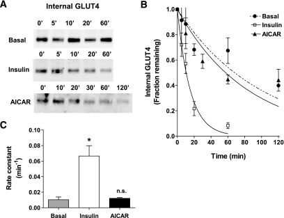
Results: In rat epitrochlearis muscle, insulin exposure leads to a sixfold stimulation of the GLUT4 exocytosis rate (with basal and insulin-stimulated rate constants of 0.010 and 0.067 min(-1), respectively). In human vastus lateralis muscle, insulin stimulates GLUT4 translocation by a similar sixfold increase in the exocytosis rate constant (with basal and insulin-stimulated rate constants of 0.011 and 0.075 min(-1), respectively). In contrast, AICAR treatment does not markedly increase exocytosis in either rat or human muscle.
Conclusions: Insulin stimulation of the GLUT4 exocytosis rate constant is sufficient to account for most of the observed increase in glucose transport activity in rat and human muscle.
Figures
Similar articles
-
Chronic treatment with 5-aminoimidazole-4-carboxamide-1-beta-D-ribofuranoside increases insulin-stimulated glucose uptake and GLUT4 translocation in rat skeletal muscles in a fiber type-specific manner.Diabetes. 2001 Jan;50(1):12-7. doi: 10.2337/diabetes.50.1.12. Diabetes. 2001. PMID: 11147776
-
Effect of insulin on GLUT4 cell surface content and turnover rate in human skeletal muscle as measured by the exofacial bis-mannose photolabeling technique.Diabetes. 1997 Dec;46(12):1965-9. doi: 10.2337/diab.46.12.1965. Diabetes. 1997. PMID: 9392481
-
Contraction stimulates translocation of glucose transporter GLUT4 in skeletal muscle through a mechanism distinct from that of insulin.Proc Natl Acad Sci U S A. 1995 Jun 20;92(13):5817-21. doi: 10.1073/pnas.92.13.5817. Proc Natl Acad Sci U S A. 1995. PMID: 7597034 Free PMC article.
-
Role of SNARE's in the GLUT4 translocation response to insulin in adipose cells and muscle.J Basic Clin Physiol Pharmacol. 1998;9(2-4):153-65. doi: 10.1515/jbcpp.1998.9.2-4.153. J Basic Clin Physiol Pharmacol. 1998. PMID: 10212832 Review.
-
Spatiotemporal Regulators for Insulin-Stimulated GLUT4 Vesicle Exocytosis.J Diabetes Res. 2017;2017:1683678. doi: 10.1155/2017/1683678. Epub 2017 Apr 25. J Diabetes Res. 2017. PMID: 28529958 Free PMC article. Review.
Cited by
-
Leptin reduces the expression and increases the phosphorylation of the negative regulators of GLUT4 traffic TBC1D1 and TBC1D4 in muscle of ob/ob mice.PLoS One. 2012;7(1):e29389. doi: 10.1371/journal.pone.0029389. Epub 2012 Jan 9. PLoS One. 2012. PMID: 22253718 Free PMC article.
-
Rab5 activity regulates GLUT4 sorting into insulin-responsive and non-insulin-responsive endosomal compartments: a potential mechanism for development of insulin resistance.Endocrinology. 2014 Sep;155(9):3315-28. doi: 10.1210/en.2013-2148. Epub 2014 Jun 16. Endocrinology. 2014. PMID: 24932807 Free PMC article.
-
Chemical biology probes of mammalian GLUT structure and function.Biochem J. 2018 Nov 20;475(22):3511-3534. doi: 10.1042/BCJ20170677. Biochem J. 2018. PMID: 30459202 Free PMC article. Review.
-
In Vitro Innervation as an Experimental Model to Study the Expression and Functions of Acetylcholinesterase and Agrin in Human Skeletal Muscle.Molecules. 2017 Aug 27;22(9):1418. doi: 10.3390/molecules22091418. Molecules. 2017. PMID: 28846617 Free PMC article. Review.
-
Reciprocal regulation of endocytosis and metabolism.Cold Spring Harb Perspect Biol. 2014 Jul 1;6(7):a016964. doi: 10.1101/cshperspect.a016964. Cold Spring Harb Perspect Biol. 2014. PMID: 24984778 Free PMC article. Review.
References
-
- Eriksson J, Koranyi L, Bourey R, Schalin-Jantti C, Widen E, Mueckler M, Permutt AM, Groop LC: Insulin resistance in type 2 (non-insulin-dependent) diabetic patients and their relatives is not associated with a defect in the expression of the insulin-responsive glucose transporter (GLUT-4) gene in human skeletal muscle. Diabetologia 35: 143– 147, 1992 - PubMed
-
- Cline GW, Petersen KF, Krssak M, Shen J, Hundal RS, Trajanoski Z, Inzucchi S, Dresner A, Rothman DL, Shulman GI: Impaired glucose transport as a cause of decreased insulin-stimulated muscle glycogen synthesis in type 2 diabetes. N Engl J Med 341: 240– 246, 1999 - PubMed
-
- Shulman GI, Rothman DL, Jue T, Stein P, Defronzo RA, Shulman RG: Quantitation of muscle glycogen synthesis in normal subjects and subjects with non-insulin-dependent diabetes by 13C nuclear magnetic resonance spectroscopy. N Engl J Med 322: 223– 228, 1990 - PubMed
-
- Kahn BB, Rosen AS, Bak JF, Andersen PH, Damsbo P, Lund S, Pedersen O: Expression of GLUT1 and GLUT4 glucose transporters in skeletal muscle of humans with insulin-dependent diabetes mellitus: regulatory effects of metabolic factors. J Clin Endocrinol Metab 74: 1101– 1109, 1992 - PubMed
Publication types
MeSH terms
Substances
Grants and funding
LinkOut - more resources
Full Text Sources
Medical
Miscellaneous
