. 2009 Apr;29(8):2139-54.
doi: 10.1128/MCB.01274-08. Epub 2009 Feb 2.

Myocilin is a modulator of Wnt signaling

Affiliations

Myocilin is a modulator of Wnt signaling

Heung-Sun Kwon et al. Mol Cell Biol. 2009 Apr.

Abstract

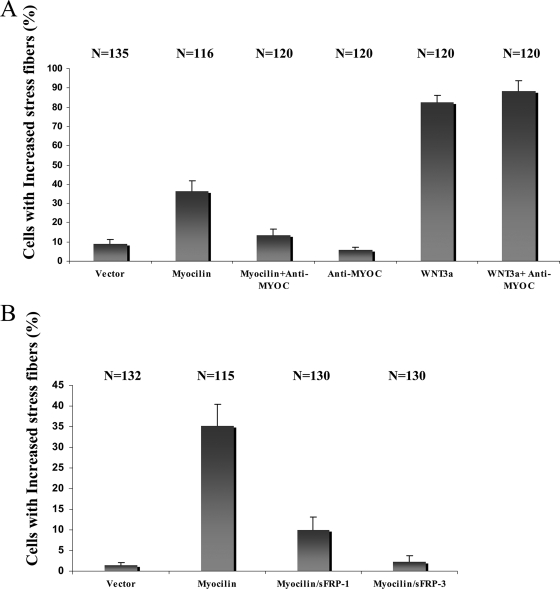
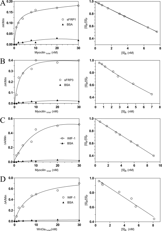
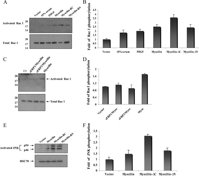
It is well documented that mutations in the MYOCILIN gene may lead to juvenile- and adult-onset primary open-angle glaucoma. However, the functions of wild-type myocilin are still not well understood. To study the functions of human myocilin and its two proteolytic fragments, these proteins were expressed in HEK293 cells. Conditioned medium from myocilin-expressing cells, as well as purified myocilin, induced the formation of stress fibers in primary cultures of human trabecular meshwork or NIH 3T3 cells. Stress fiber-inducing activity of myocilin was blocked by antibodies against myocilin, as well as secreted inhibitors of Wnt signaling, secreted Frizzled-related protein 1 (sFRP1) or sFRP3, and beta-catenin small interfering RNA. Interaction of myocilin with sFRP1, sFRP3, and several Frizzled receptors was confirmed by immunoprecipitation experiments and by binding of myocilin to the surface of cells expressing cysteine-rich domains of different Frizzled and sFRPs. Treatment of NIH 3T3 cells with myocilin and its fragments induced intracellular redistribution of beta-catenin and its accumulation on the cellular membrane but did not induce nuclear accumulation of beta-catenin. Overexpression of myocilin in the eye angle tissues of transgenic mice stimulated accumulation of beta-catenin in these tissues. Myocilin and Wnt proteins may perform redundant functions in the mammalian eye, since myocilin modulates Wnt signaling by interacting with components of this signaling pathway.


Figures

FIG. 1.
FIG. 1.
Schematic diagram of myocilin constructs that were used in the present study (A), secretion of different myocilin isoforms (B), and SDS-PAGE analysis of purified myocilin proteins (C). (A) A signal peptide and olfactomedin domain are shown in red and dark blue, respectively. The FLAG epitope is shown in brown. The arrow marks the proteolytic cleavage site. Numbers at the top indicate corresponding positions in the myocilin amino acid sequence. (B) HEK293 cells were transiently transfected with a construct encoding full-length human myocilin, and the presence of different myocilin forms in conditioned medium (lanes 2 and 3) was analyzed 48 h after transfection by Western blotting with antibodies to the N-terminal part of myocilin (lane 2) or FLAG epitope (lane 3). A total of 5 μg of total cell extract was stained with antibodies to the N-terminal part of myocilin (lane 1). (C) A total of 0.5 μg of indicated purified proteins was separated by SDS-4 to 12% PAGE and stained with Coomassie blue.
FIG. 2.
FIG. 2.
Myocilin-induced formation of actin stress fibers in human trabecular meshwork cells. Cells were treated with 1 μg of the indicated myocilin proteins/ml for 1 h and then stained with rhodamine phalloidin. (B) Quantification of studies corresponding to panel A. The number of cells evaluated is shown at the top of each bar. These experiments were performed three times. Scale bar, 10 μm.
FIG. 3.
FIG. 3.
Myocilin-induced formation of actin stress fibers in NIH 3T3 cells. (A) Cells were treated with CM from HEK293 cells transiently transfected with indicated plasmids for 4 h and then stained with rhodamine phalloidin. Representative fields are shown. (B) Quantification of a typical experiment showing the percentage of cells containing actin stress fibers. The number of cells evaluated is shown at the top of each bar. These experiments were performed four times. Scale bar, 10 μm.
FIG. 4.
FIG. 4.
Effects of myocilin antiserum (A) and sFRPs (B) on actin stress fibers formation in NIH 3T3 cells. Myocilin antiserum at 10 μg/ml (A) or the indicated sFRPs at 10 μg/ml (B) was preincubated with myocilin CM for 30 min before addition to NIH 3T3 cells. The number of cells evaluated is shown at the top of each bar. These experiments were performed twice.
FIG. 5.
FIG. 5.
Changes in subcellular localization of β-catenin in response to myocilin treatment and effects of β-catenin inhibition on stress fiber formation. NIH 3T3 cells were treated with CM from cells expressing no myocilin (A), myocilin (B), myocilin-ΔC (C), or myocilin-ΔN for 3 h and stained with antibodies to β-catenin. Note the membrane localization of β-catenin in panels B to D. These experiments were performed four times. (E) Changes in the levels of total β-catenin in CHO cells treated with CM from Wnt3a- or myocilin-expressing cells. (F) Changes in the levels of total β-catenin in NIH 3T3 cells treated with β-catenin siRNA (10, 20, 40, and 80 nM) as described in Materials and Methods. (G) Reduction of stress fiber formation in NIH 3T3 cells after reduction of β-catenin level by 80 nM siRNA. Scale bar, 10 μm.
FIG. 6.
FIG. 6.
Upregulation of β-catenin in the eyes of 20-month-old wild-type and transgenic mice. Paraffin sections of control (A) and transgenic (B) eyes were stained with antibodies to β-catenin (1:100 dilution) and DAPI. Three pairs of animals were analyzed. A typical staining pattern is shown. cb, ciliary body; i, iris; tm, trabecular meshwork. (C) Western blot analysis of β-catenin expression in the eye angle tissues and sclera of wild-type and transgenic mice. (D) Quantification of the results shown in panel B. WT, wild type; AG, eye angle tissues; SL, sclera.
FIG. 7.
FIG. 7.
Binding of myocilin-AP to the CRDs of different Fzds and sFRPs. Myocilin-AP CM was incubated with live HEK293 cells transfected with the indicated CRD-Myc-GPI constructs. The AP activity was visualized by staining with nitro blue tetrazolium/BCIP. AP-Norrin CM was used as a control. These experiments were repeated three times. The results of a typical experiment are shown.
FIG. 8.
FIG. 8.
Physical interaction of myocilin with Fzd receptors and antagonists of Wnt signaling. HEK293 cells were cotransfected with the indicated constructs (A and D). Mouse heart extracts were used in panels B and C. CM was collected 48 h after transfection in the case of secreted antagonists and treated with Myc antibodies (A). (B and C) Heart extracts were immunoprecipitated with antibodies to mouse myocilin. After immunoprecipitation, proteins were eluted from the beads, separated by SDS-PAGE, and then probed with antibodies to sFRP3 (B) or sFRP1 (C). (D) HEK293 cells were lysed, immunoprecipitated with anti-FLAG beads, and analyzed as described above with antibodies to HA for detection. Shown are Western blots of CM (A, right) or cell lysates (B to D, lower panels) before immunoprecipitation probed with the indicated antibodies. These experiments were repeated more than two times.
FIG. 9.
FIG. 9.
Binding of myocilin to sFRP1, sFRP3, WIF-1, and BSA (left panels). The binding of Wnt3a to WIF-1 and BSA is shown for comparison. The vertical axis shows the increase in absorbance at 405 nm. The right panels represent Scatchard plots of the data for myocilin binding to corresponding proteins. The vertical axis shows the ratio of bound to free myocilin; the horizontal axis shows the concentration of bound myocilin. These experiments were repeated three times.
FIG. 10.
FIG. 10.
Physical interaction of myocilin and WIF-1. HEK293 cells were cotransfected with the indicated constructs. CM was collected 48 h after transfection, and protein complexes were precipitated with FLAG antibody beads. Proteins eluted from the beads were separated by SDS-PAGE and probed with peroxidase-conjugated antibodies to human IgG (upper panel). The lower panel shows a Western blot of the CM before immunoprecipitation probed with anti-FLAG antibodies. These experiments were repeated two times.
FIG. 11.
FIG. 11.
Myocilin activates Rac1 and JNK. The indicated myocilin CMs were added to NIH 3T3 cells for 30 min, and the levels of activated Rac1 (A) and JNK (E) were measured. The ratio between activated and total Rac1 was about 1:100 for myocilin-ΔC. Preincubation of CM with sFRP1 or sFRP3 eliminated activation of Rac1 by myocilin (C). Panels B, D, and F represent quantification of the results shown in panels A, C, and E, respectively. These experiments were repeated at least twice.
FIG. 12.
FIG. 12.
Schematic diagram of myocilin action. Myocilin may bind Wnt antagonists WIF-1 and sFRPs and compete with Wnt for binding to several Fzd receptors. Proteins that are affected by myocilin treatment are indicated in red. Thin uninterrupted lines with two arrows indicate proteins that interact with each other.

References

    1. Adam, M. F., A. Belmouden, P. Binisti, A. P. Brezin, F. Valtot, A. Bevhetoille, J.-C. Dascotte, B. Copin, L. Gomez, A. Chaventre, J.-F. Bach, and H.-J. Garchon. 1997. Recurrent mutations in a single exon encoding the evolutionary conserved olfactomedin-homology domain of TIGR in familial open-angle glaucoma. Hum. Mol. Genet. 62091-2097. - PubMed
    1. Alward, W. L., Y. H. Kwon, C. L. Khanna, A. T. Johnson, S. S. Hayreh, M. B. Zimmerman, J. Narkiewicz, J. L. Andorf, P. A. Moore, J. H. Fingert, V. C. Sheffield, and E. M. Stone. 2002. Variations in the myocilin gene in patients with open-angle glaucoma. Arch. Ophthalmol. 1201189-1197. - PubMed
    1. Aroca-Aguilar, J. D., F. Sanchez-Sanchez, S. Ghosh, M. Coca-Prados, and J. Escribano. 2005. Myocilin mutations causing glaucoma inhibit the intracellular endoproteolytic cleavage of myocilin between amino acids Arg226 and Ile227. J. Biol. Chem. 28021043-21051. - PubMed
    1. Barembaum, M., T. A. Moreno, C. LaBonne, J. Sechrist, and M. Bronner-Fraser. 2000. Noelin-1 is a secreted glycoprotein involved in generation of the neural crest. Nature Cell Biol. 2219-225. - PubMed
    1. Barker, N., and H. Clevers. 2006. Mining the Wnt pathway for cancer therapeutics. Nat. Rev. Drug Discov. 5997-1014. - PubMed

Publication types

LinkOut - more resources