. 2009 Feb 17;106(7):2418-22.
doi: 10.1073/pnas.0811400106. Epub 2009 Feb 2.

Melanin-concentrating hormone neurons discharge in a reciprocal manner to orexin neurons across the sleep-wake cycle

Affiliations

Melanin-concentrating hormone neurons discharge in a reciprocal manner to orexin neurons across the sleep-wake cycle

Oum Kaltoum Hassani et al. Proc Natl Acad Sci U S A. .

Abstract

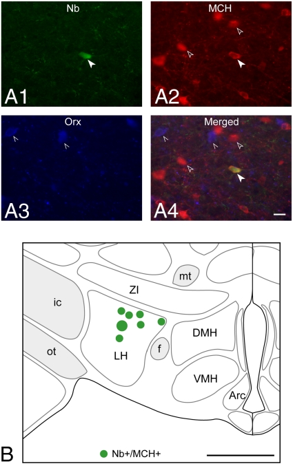
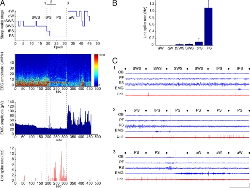
Neurons containing melanin-concentrating hormone (MCH) are codistributed with neurons containing orexin (Orx or hypocretin) in the lateral hypothalamus, a peptide and region known to be critical for maintaining wakefulness. Evidence from knockout and c-Fos studies suggests, however, that the MCH neurons might play a different role than Orx neurons in regulating activity and sleep-wake states. To examine this possibility, neurons were recorded across natural sleep-wake states in head-fixed rats and labeled by using the juxtacellular technique for subsequent immunohistochemical identification. Neurons identified as MCH+ did not fire during wake (W); they fired selectively during sleep, occasionally during slow wave sleep (SWS) and maximally during paradoxical sleep (PS). As W-Off/Sleep-On, the MCH neurons discharged in a reciprocal manner to the W-On/Sleep-Off Orx neurons and could accordingly play a complementary role to Orx neurons in sleep-wake state regulation and contribute to the pathophysiology of certain sleep disorders, such as narcolepsy with cataplexy.


Conflict of interest statement

The authors declare no conflict of interest.

Figures

Fig. 1.
Fig. 1.
Recorded, Nb-labeled, MCH-immunopositive neurons were located near the Orx neurons in the lateral hypothalamus. (A) Fluorescence image of Nb-labeled (green, Cy2, filled arrowhead in A1) neuron (c134u01), which was immunopositive for MCH (red, Cy3, arrowheads in A2) and located near codistributed Orx+ neurons (blue, Cy5, pointers in A3 and in merged image in A4). (Scale bar, 25 μm.) (B) Location of recorded, Nb+/MCH+ neurons (n = 7) on a coronal atlas section (≈ −2.8 mm from bregma or 6.6 mm anterior to interaural zero) through the lateral hypothalamus. The cell shown in A is represented as the largest symbol in B. (Scale bar, 1 mm.) Arc, arcuate nucleus; DMH, dorsomedial hypothalamic nucleus; f, fornix; ic, internal capsule; LH, lateral hypothalamus; mt, mammillo-thalamic tract; ot, optic tract; VMH, ventromedial hypothalamic nucleus; ZI, zona incerta.
Fig. 2.
Fig. 2.
Nb+/MCH+ neurons discharged only during sleep. (A) Data from Nb+/MCH+ unit (c134u01, Fig. 1) showing the sleep–wake stage scored per 10-s epoch, together with simultaneous EEG amplitude (μV/Hz with frequency on y axis and amplitude scaled according to color), EMG amplitude (μV), and unit spike rate (Hz) per second of the recording session. Note that the unit fired during SWS or tSWS, tPS, and PS when EEG showed prominent slow (<5 Hz) or theta (≈6–8 Hz) activity and EMG amplitude was relatively low. Vertical dashed lines indicate precise moments of transition between SWS and tPS, tPS and PS, and PS and aW here and within corresponding segments 1, 2, and 3 in C. (B) Mean spike rate (Hz) of the Nb+/MCH+ unit per sleep–wake stage. Note that it did not fire during aW or qW, fired very little during tSWS, SWS, and tPS, and fired maximally during PS. (C) Polygraphic records (from 1-min periods indicated in A) of the unit discharge along with EEG and EMG activity during passages between states: (1) from SWS to tPS, (2) from tPS to PS, and (3) from PS to aW. Note that the unit fired only very occasionally with 1 or 2 spikes during periods of SWS and tPS (shown in C1). It discharged phasically in groups of spikes to reach its highest rates during PS (shown in C2). It ceased firing on arousal (marked by dashed line indicating transition during uncategorized epoch) and during the subsequent aW period (shown in C3). Movies S1–S3 are also shown of 30-s segments of continuous SWS (1), PS (2), and aW (3) taken before (1) or after (2 and 3) the transitional periods shown in C. Calibrations: horizontal, 10 s; vertical, 1 mV (EEG, EMG), 2 mV (unit). OB, olfactory bulb; PF, prefrontal cortex; RS, retrosplenial cortex.
Fig. 3.
Fig. 3.
MCH neurons discharged in a reciprocal manner to Orx neurons across sleep–wake states. (A) The mean spike rate (Hz) per stage of Nb+/Orx+ units (n = 6; from ref. 15) varies in a reciprocal manner to that of Nb+/MCH+ units (n = 6; this study). (B) The reciprocal firing profiles were not correlated with EEG gamma or delta activity but were correlated in an inverse manner with EMG amplitude, positively for the wake-active Orx neurons and negatively for the MCH sleep-active neurons. EEG and EMG plotted as mean normalized amplitude from the same recordings as units.

References

    1. Jones BE. In: Principles and Practice of Sleep Medicine. Kryger MH, Roth T, Dement WC, editors. Philadelphia: Elsevier Saunders; 2005. pp. 136–153.
    1. Peyron C, et al. Neurons containing hypocretin (orexin) project to multiple neuronal systems. J Neurosci. 1998;18:9996–10015. - PMC - PubMed
    1. Chemelli RM, et al. Narcolepsy in orexin knockout mice: Molecular genetics of sleep regulation. Cell. 1999;98:437–451. - PubMed
    1. Lin L, et al. The sleep disorder canine narcolepsy is caused by a mutation in the hypocretin (orexin) receptor 2 gene. Cell. 1999;98:365–376. - PubMed
    1. Thannickal TC, et al. Reduced number of hypocretin neurons in human narcolepsy. Neuron. 2000;27:469–474. - PMC - PubMed

Publication types