. 2009 Mar;119(3):611-23.
doi: 10.1172/JCI35977. Epub 2009 Feb 2.

Antioxidant or neurotrophic factor treatment preserves function in a mouse model of neovascularization-associated oxidative stress

Affiliations

Antioxidant or neurotrophic factor treatment preserves function in a mouse model of neovascularization-associated oxidative stress

Michael I Dorrell et al. J Clin Invest. 2009 Mar.

Abstract

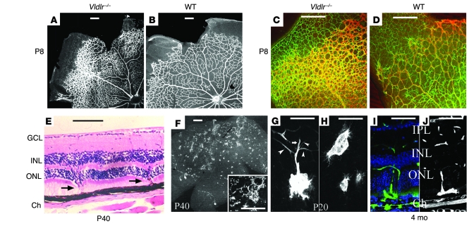
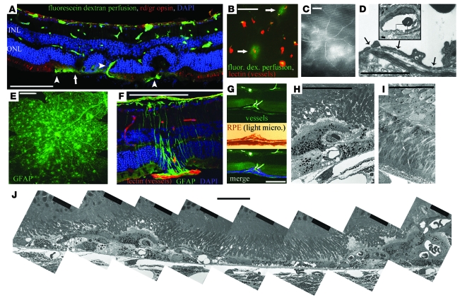
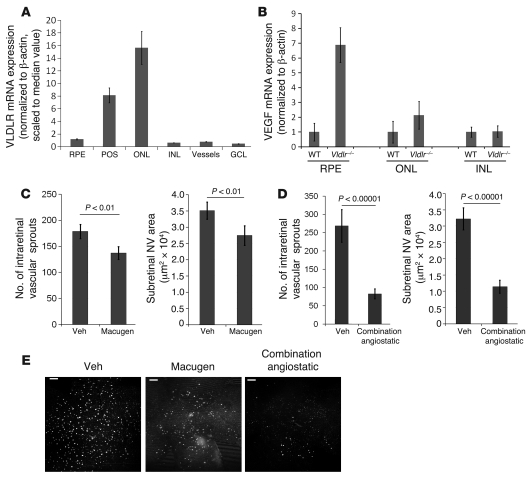
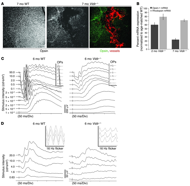
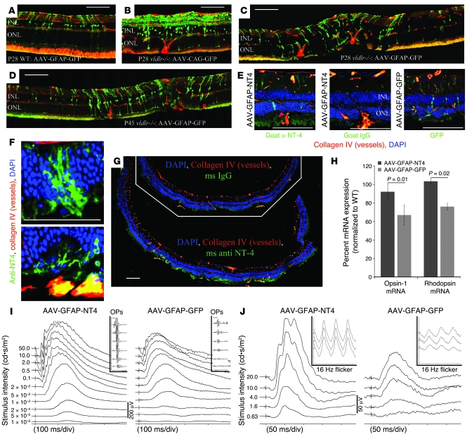
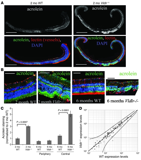
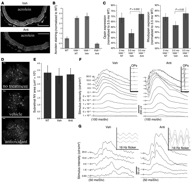
In several disease states, abnormal growth of blood vessels is associated with local neuronal degeneration. This is particularly true in ocular diseases such as retinal angiomatous proliferation (RAP) and macular telangiectasia (MacTel), in which, despite the absence of large-scale leakage or hemorrhage, abnormal neovascularization (NV) is associated with local neuronal dysfunction. We describe here a retinal phenotype in mice with dysfunctional receptors for VLDL (Vldlr-/- mice) that closely resembles human retinal diseases in which abnormal intra- and subretinal NV is associated with photoreceptor cell death. Such cell death was evidenced by decreased cone and, to a lesser extent, rod opsin expression and abnormal electroretinograms. Cell death in the region of intraretinal vascular abnormalities was associated with an increased presence of markers associated with oxidative stress. Oral antioxidant supplementation protected against photoreceptor degeneration and preserved retinal function, despite the continued presence of abnormal intra- and subretinal vessels. What we believe to be novel, Müller cell-based, virally mediated delivery of neurotrophic compounds specifically to sites of NV was also neuroprotective. These observations demonstrate that neuronal loss secondary to NV can be prevented by the use of simple antioxidant dietary measures or cell-based delivery of neurotrophic factors, even when the underlying vascular phenotype is not altered.


Figures

Figure 1
Figure 1. Intraretinal vascular abnormalities in Vldlr–/– mouse retinas.
(A and B) The developing superficial vascular plexus of Vldlr–/– retinas was hyperdense, particularly in the periphery, at P8. (C and D) Peripheral hypervascularity (isolectin GS; red) was associated with increased density of astrocytes (GFAP; green). (E) H&E-stained retinal sections from a P40 Vldlr–/– mouse showed intraretinal vessels (arrows) that originated from the inner retinal vascular plexuses and migrated through the photoreceptors to the subretinal space. GCL, ganglion cell layer; Ch, choroid. (F) These intraretinal vessels formed retinal-retinal anastomoses throughout the central two-thirds of the Vldlr–/– retinas. Inset shows a higher-magnification view. (G) Intraretinal vessels originated from multiple sprouts off the deep (arrowheads) and intermediate vascular plexuses (arrow) and could form large angiomatous structures within the subretinal space. (H) Filopodia extended from subretinal vascular endothelial cells and extended laterally to form retinal vascular anastomoses. (I and J) Fluorescein angiography (I) or isolectin GS staining (J) demonstrated the retinal origin of the intraretinal and subretinal NV. IPL, inner plexiform layer. Scale bars: 100 μm.
Figure 2
Figure 2. Characterization of abnormal NV in the Vldlr–/– mouse retina.
(A) Retinal cross-section montage demonstrating multiple retinal abnormalities in the Vldlr–/– mouse retina at 3 months of age, including subretinal vessels (arrowheads), vascular leak (arrow), abnormal retinal morphology, abnormal neuronal layering in the ONL and INL, and loss of red/green (rd/gr) cone opsin staining. (B and C) Vascular leak was associated with a small subset of subretinal vessels, as demonstrated by extravasation of perfused 43-kDa fluorescein-labeled dextran. (D) Fenestrae within endothelial cells of the subretinal vessels were observed by electron microscopy. Inset shows a lower-magnification view, in which the boxed region defines the bounds of the higher-magnification view. (E) Punctate GFAP staining in retinal whole mounts demonstrated glial activation associated with abnormal NV in the Vldlr–/– mouse retinas. (F) This punctate staining represented Müller cells specifically activated around the abnormal intraretinal vessels. (G and H) The RPE formed multicellular layers that engulfed abnormal intraretinal vessels. (I) Electron microscopy demonstrated normal retinal morphology at P12, just prior to NV onset. (J) Montage of multiple electron microscopy images showing retinal damage spatially associated with abnormal NV in 2 separate regions, while intermittent retinal morphology between lesions remained mostly normal. Scale bars: 100 μm (AC and EG); 50 μm (HJ); 10 μm (D).
Figure 3
Figure 3. VEGF upregulation is associated with intraretinal NV in Vldlr–/– mice.
(A) Most VLDLR expression in laser-captured retinal tissue from P14 C57BL6/J mice occurred in the ONL and photoreceptor segments (POS), with lower expression in the RPE and other retinal tissues (triplicate data). (B) In Vldlr–/– retinas, significant upregulation of VEGF occurred within the RPE (P < 0.0001) and photoreceptors (P < 0.05) at P14 (triplicate data). (C) Blocking VEGF165 activity with intravitreal Macugen injection at P12 significantly reduced the number of intraretinal NV sprouts and the area of subretinal NV at P20 in Vldlr–/– retinas. n = 10–12 retinas per group. Veh, vehicle. (D) Combination angiostatic therapy reduced abnormal retinal NV to an even greater extent. n = 10–12 retinas per group. (E) Whole retina images of vessels, focused within the subretinal space, showed a sharp reduction in intraretinal NV after treatment with Macugen and combination angiostatics. Scale bars: 250 μm. Error bars denote SEM.
Figure 4
Figure 4. Photoreceptor degeneration, loss of retinal function, and increased oxidative stress are associated with intraretinal NV in Vldlr–/– mouse retinas.
(A) Opsin staining was reduced in Vldlr–/– retinal whole mounts, with loss of opsin directly associated with the presence of subretinal NV. Original magnification, ×100. (B) Reduced mRNA production of opsin-1 (cone-specific opsin) and rhodopsin (rod-specific opsin) in retinas of 2- and 7-mo-old Vldlr–/– mice compared with age-matched C57BL6/J WT controls, indicating a substantial loss of cones and, to a lesser extent, rods. Error bars denote SEM. n = 18 [2 mo WT], 28 [2 mo Vldlr–/–], 36 [7 mo WT], 30 [7 mo Vldlr–/–]. (C) Photoreceptor degeneration resulted in abnormal ERGs, characterized by delayed responses in scotopic measurements and dramatic reductions in the OPs (insets). (D) Photopic ERGs of Vldlr–/– mice were also characterized by reduced and delayed responses, with particularly reduced responses to light flicker (insets), a specific measurement of cone activity.
Figure 5
Figure 5. Targeted delivery of NT4 to sites of subretinal NV protects Vldlr–/– retinas from neuronal degeneration.
(A) AAV-GFAP-GFP–treated WT retinas exhibited minimal GFP expression in the inner retina and no expression in the outer retina. (B) AAV-CAG-GFP caused nonspecific GFP expression, mainly limited to the inner retina. (C and D) At 2 wk after intravitreal AAV-GFAP-GFP injection, GFP was observed in Müller glia specifically surrounding subretinal NV in P28 Vldlr–/– mouse retinas (C), which was maintained at P45, 1 mo after injection (D). (EG) NT4 gene product was produced in Müller cells adjacent to subretinal NV in AAV-GFAP-NT4–treated Vldlr–/– retinas (E and F), resulting in accumulation of NT4 at photoreceptor segments (G). Line in G separates control, pre–immune IgG–stained retina (top) from retina stained with anti-NT4 antibodies (bottom). (H) Quantitative RT-PCR analysis of opsin-1 and rhodopsin mRNA expression, normalized to age-matched WT controls, demonstrated protective effects of AAV-GFAP-NT4 treatment. Error bars denote SEM. (I and J) ERG analysis demonstrated that AAV-GFAP-NT4 treatment attenuated loss of retinal function. Scale bars: 100 μm (AE and G); 50 μm (F).
Figure 6
Figure 6. Evidence of oxidative stress associated with subretinal NV in Vldlr–/– mouse retinas.
(A) Acrolein staining increased in Vldlr–/– retinas compared with age-matched WT controls. Acrolein staining in Vldlr–/– retinas was largely within the central retina, where intraretinal NV is most prominent. Panels are composite montages of multiple serial micrographs. (B) Higher-magnification images demonstrating that acrolein staining localized to the photoreceptor layer and INL. (C) Acrolein staining significantly increased in 2-mo-old Vldlr–/– compared with WT retinas. At 6 mo of age, acrolein staining was similar in the peripheral retinas of WT and Vldlr–/– mice (P = 0.245), but was significantly stronger in the central regions of Vldlr–/– retinas, where subretinal NV occurs, compared with retinas of WT mice. Error bars denote SEM. (D) Expression of genes involved in oxidative stress defense mechanisms was similar between P21 Vldlr–/– and WT retinas. Solid line indicates equivalent expression levels; dotted lines represent 95% confidence level ranges. Scale bars: 500 μm (A); 50 μm (B).
Figure 7
Figure 7. Antioxidants protect retinas from degeneration and reduced function in Vldlr–/– retinas.
(A and B) In retinas of antioxidant-treated 2-mo-old Vldlr–/– mice, decreased acrolein staining was observed compared with age-matched vehicle-treated controls. Panels in A are composite montages of multiple serial micrographs. Error bars denote SEM. NT, not treated; Anti, antioxidant treatment. (CG) Vldlr–/– mice were subjected to a 6-wk treatment with vehicle or antioxidants, beginning at 2 mo of age. (C) Antioxidant treatment attenuated the loss of cones and rods, as demonstrated by normalized expression of opsin-1 and rhodopsin, respectively. n = 12 retinas per group. Error bars denote SEM. (D and E) The extent of subretinal NV in 3.5-mo-old Vldlr–/– mouse retinas was not affected by antioxidant treatment. n = 12 retinas per group. Error bars denote SEM. (F and G) ERGs were normalized after 6 wk of antioxidant treatment. Note decreased and delayed signaling in vehicle-treated Vldlr–/– mice compared with those treated with antioxidants. Scale bars: 250 μm.

References

    1. Barcia C., Emborg M.E., Hirsch E.C., Herrero M.T. Blood vessels and parkinsonism. Front. Biosci. 2004;9:277–282. doi: 10.2741/1145. - DOI - PubMed
    1. Browne S.E., Beal M.F. Oxidative damage in Huntington’s disease pathogenesis. Antioxid. Redox Signal. 2006;8:2061–2073. doi: 10.1089/ars.2006.8.2061. - DOI - PubMed
    1. Nunomura A., et al. Involvement of oxidative stress in Alzheimer disease. J. Neuropathol. Exp. Neurol. 2006;65:631–641. doi: 10.1097/01.jnen.0000228136.58062.bf. - DOI - PubMed
    1. Zhu X., et al. Vascular oxidative stress in Alzheimer disease. J. Neurol. Sci. 2007;257:240–246. doi: 10.1016/j.jns.2007.01.039. - DOI - PMC - PubMed
    1. Faucheux B.A., Bonnet A.M., Agid Y., Hirsch E.C. Blood vessels change in the mesencephalon of patients with Parkinson’s disease. Lancet. 1999;353:981–982. - PubMed

Publication types

MeSH terms