Review
. 2009 Apr;87(4):363-74.
doi: 10.1007/s00109-009-0440-9. Epub 2009 Feb 3.

Tumor initiating cells in malignant gliomas: biology and implications for therapy

Affiliations
Review

Tumor initiating cells in malignant gliomas: biology and implications for therapy

Costas G Hadjipanayis et al. J Mol Med (Berl). 2009 Apr.

Abstract

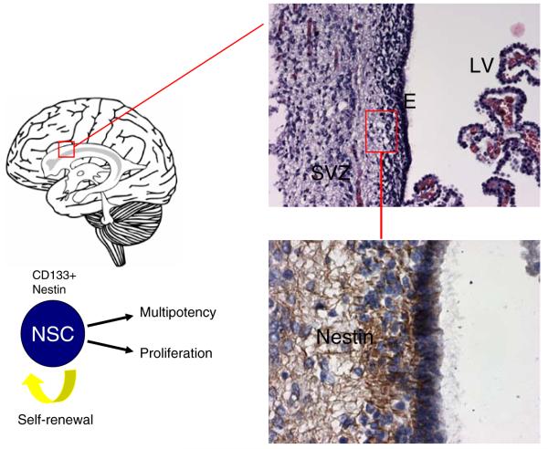
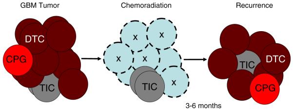
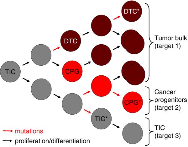
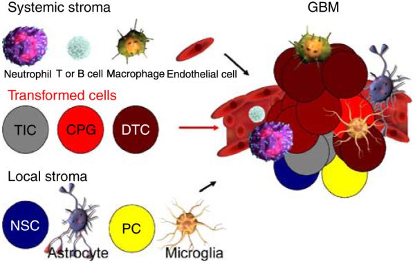
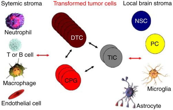
A rare subpopulation of cells within malignant gliomas, which shares canonical properties with neural stem cells (NSCs), may be integral to glial tumor development and perpetuation. These cells, also known as tumor initiating cells (TICs), have the ability to self-renew, develop into any cell in the overall tumor population (multipotency), and proliferate. A defining property of TICs is their ability to initiate new tumors in immunocompromised mice with high efficiency. Mounting evidence suggests that TICs originate from the transformation of NSCs and their progenitors. New findings show that TICs may be more resistant to chemotherapy and radiation than the bulk of tumor cells, thereby permitting recurrent tumor formation and accounting for the failure of conventional therapies. The development of new therapeutic strategies selectively targeting TICs while sparing NSCs may provide for more effective treatment of malignant gliomas.


Figures

Fig. 1
Fig. 1
Source of neural stem cells and GBM tumors. The subventricular zone (SVZ) is found beneath the ependymal (E) layer that lines the lateral ventricles (LV) of the brain and is believed to be the origin of GBM tumors (upper section). Neural stem cells (NSCs) originate from this area of the brain and are characterized by the cell surface markers CD133 and nestin. Immunohistochemistry staining of the SVZ shows the presence of nestin-expressing cells, some of which represent NSCs (lower section). The structures on the right side of the upper section are choroid plexus protrusions from which the CSF filling the ventricles originates
Fig. 2
Fig. 2
Possible lineage relationships for the ontogeny and production of tumor-initiating cells (TICs) and generation of GBM tumors. During normal CNS differentiation neural stem cells (NSCs) undergo an amplification step to produce transit-amplifying progenitor cells (TACs), which then differentiate into committed progenitor cells (PCs*). PC* (neuronal or glial) retain the capacity of producing progeny along either neuronal or glial lineages (oligodendrocytes and/or astrocytes), but not both. Mutations generating gliomas can occur at all levels within this lineage and produce TICs or differentiated tumor cells (DTCs). TICs are thought to be stem-like in behavior by their ability to self-renew, proliferate, and generate DTCs or cancer progenitor cells (CPG) within the tumor mass. While this model suggests unilateral progression toward mutated cell populations that become terminally differentiated cancer cells, in reality, there is evidence in the literature for more plasticity, and de-differentiation events may also take place to generate self-propagating cancer cells from astrocytes and oligodendrocytes. It is unknown whether neurons can generate cancer cells
Fig. 3
Fig. 3
TICs as the origin of GBM tumor resistance to therapy. Recurrence of malignant brain tumors in 3-6 months may be related to the resistance of TICs to standard therapies, such as chemoradiation, and their ability to generate new tumors. Note that the bulk of the tumor (shown by empty circles marked by X) responds to therapy, but the TICs are unaffected and regenerate the tumor. It is currently unknown whether CPGs may possess the capacity to regenerate the tumor as well. Clearly, this is a simplified model, and different cell populations in the tumor will respond differentially to individual therapies, depending on their intrinsic resistance mechanisms; these may include expression of drug transporters, genetic composition, dependence upon specific signaling pathways, aerobic and anaerobic metabolism, and ability to cope with reactive oxygen species (ROS) stress. TIC tumor-initiating cell, CPG cancer progenitor cell, DTC differentiated tumor cell
Fig. 4
Fig. 4
Therapeutic intervention for CNS tumors requires targeting of multiple cell types. Treatments for CNS tumors may require targeting of differentiated tumor cells (DTC), which likely make up the bulk of the tumor (target 1), cancer progenitor cells (CPG; target 2), and TICs (target 3). TICs can give rise to DTCs through mutational events, while TICs may develop CPGs by mutations or differentiation events. CPGs are likely committed to the development of DTCs after proliferation and differentiation. Each of these cell populations may require distinct therapies, either delivered simultaneously or sequentially. Clonal evolution of these cell populations in the tumor is linked to the sequential accumulation of genetic defects (mutations), which will drive cell expansion. Activation of differentiation programs will limit the pluripotency of certain cells but not necessarily their replicative potential. It is currently unknown whether DTC can undergo multiple rounds of cell division or are rapidly terminally differentiated and cease proliferation. This simplified schematic does not exclude the existence of a whole hierarchy of TIC, CPG, and DTC with multiple genetic defects (asterisk). It also allows for the coexistence of different cell populations, which may share initial genetic defects but have distinct downstream mutations. Conceivably, such cell populations may even gain a mutual benefit by providing reciprocal cell survival signals. TIC tumor-initiating cell, CPG cancer progenitor cell, DTC differentiated tumor cell
Fig. 5
Fig. 5
Heterogeneous composition of GBM including vascular niche and other cells. The cellular complexity of the tumor is shown, highlighting the fact that any therapy will have to take into consideration all cellular elements of this complex tissue, which likely functions as a self-sustaining and expanding cancer organ growing as a “parasite” of the normal host. Systemic stromal cells (neutrophils, T or B cells, macrophages, and endothelial cells) and local stromal cells (NSCs, astrocytes, PCs, and microglia) are shown to potentially interact with the transformed cells (TICs, CPGs, and DTCs) to form the GBM tumor. The vascular niche provided by endothelial cells is a well-established component to the maintenance and survival of TICs and GBM tumors. It will be the key to determine which stromal cell populations (systemic or local) contribute to tumor initiation and progression and what extracellular signals regulate their recruitment to the tumor. Antagonizing such signals, possibly in the systemic blood circulation, may be a novel approach to therapy. NSC neural stem cell, PC progenitor cell, TIC tumor-initiating cell, CPG cancer progenitor cell, DTC differentiated tumor cell
Fig. 6
Fig. 6
Hypothetical interactions between tumor cells and stromal cells. The complexity of heterotypic cell-cell communication in GBM needs extensive study. Are critical signals present which sustain and help the growth of the different tumor cell populations (TIC, CPG, and DTC)? How do these interact with stromal cells, either from the local brain microenvironment or recruited systemically, and are such signals essential for tumor maintenance, progression, and recurrence? Is there transfer of oncogenic proteins from brain cancer cells to normal stroma through secreted vesicles, e.g., oncosomes? Are there tumor suppressor effects from normal stroma onto cancer cells that may be amplified for therapy? TIC tumor-initiating cell, CPG cancer progenitor cell, DTC differentiated tumor cell, NSC neural stem cell, PC progenitor cell

References

    1. Jordan CT, Guzman ML, Noble M. Cancer stem cells. N Engl J Med. 2006;355:1253–1261. - PubMed
    1. Reya T, Morrison SJ, Clarke MF, Weissman IL. Stem cells, cancer, and cancer stem cells. Nature. 2001;414:105–111. - PubMed
    1. Al-Hajj M, Becker MW, Wicha M, Weissman I, Clarke MF. Therapeutic implications of cancer stem cells. Curr Opin Genet Dev. 2004;14:43–47. - PubMed
    1. Wicha MS, Liu S, Dontu G. Cancer stem cells: an old idea—a paradigm shift. Cancer Res. 2006;66:1883–1890. discussion 1895-1886. - PubMed
    1. Lapidot T, Sirard C, Vormoor J, Murdoch B, Hoang T, Caceres-Cortes J, Minden M, Paterson B, Caligiuri MA, Dick JE. A cell initiating human acute myeloid leukaemia after transplantation into SCID mice. Nature. 1994;367:645–648. - PubMed

Publication types

LinkOut - more resources