

The CDK domain of p21 is a suppressor of IL-1beta-mediated inflammation in activated macrophages
- PMID: 19189309
- PMCID: PMC2734089
- DOI: 10.1002/eji.200838683
The CDK domain of p21 is a suppressor of IL-1beta-mediated inflammation in activated macrophages
Abstract
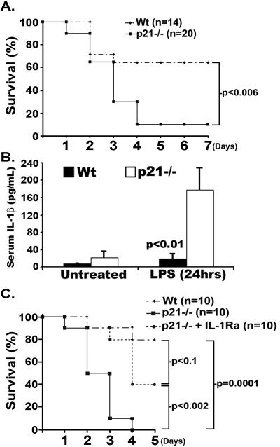
Significant morbidity and mortality can be attributed to inflammatory diseases; therefore, a greater understanding of the mechanisms involved in the progression of inflammation is crucial. Here, we demonstrate that p21((WAF1/CIP1)), an established suppressor of cell cycle progression, is a inhibitor of IL-1beta synthesis in macrophages. Mice deficient in p21 (p21(-/-)) display increased susceptibility to endotoxic shock, which is associated with increased serum levels of IL-1beta. Administration of IL-1 receptor antagonist reduces LPS-induced lethality in p21(-/-) mice. Analysis of isolated macrophages, which are one of the central producers of IL-1beta, reveals that deficiency for p21 led to more IL-1beta mRNA and pro-protein synthesis following TLR ligation. The increase in IL-1beta pro-protein is associated with elevated secretion of active IL-1beta by p21(-/-) macrophages. siRNA-mediated knockdown of p21 in human macrophages results in increased IL-1beta secretion as well. A peptide mapping strategy shows that the cyclin-dependent-kinase (CDK)-binding domain of p21 is sufficient to reduce the secretion of IL-1beta by p21(-/-) macrophages. These data suggest a novel role for p21 and specifically for the CDK-binding domain of p21((WAF1/CIP1)) in inhibiting inflammation.
Figures


References
-
- Dinarello CA. Interleukin-1beta. Crit Care Med. 2005;33:S460–462. - PubMed
-
- Furst DE, Breedveld FC, Kalden JR, Smolen JS, Burmester GR, Bijlsma JWJ, Dougados M, et al. Updated consensus statement on biological agents, specifically tumour necrosis factor {alpha} (TNF{alpha}) blocking agents and interleukin-1 receptor antagonist (IL-1ra), for the treatment of rheumatic diseases, 2005. Ann Rheum Dis. 2005;64:iv2–14. - PMC - PubMed
-
- Callen JP. Complications and adverse reactions in the use of newer biologic agents. Semin Cutan Med Surg. 2007;26:6–14. - PubMed
-
- Sherr CJ, Roberts JM. CDK inhibitors: positive and negative regulators of G1-phase progression. Genes Dev. 1999;13:1501–1512. - PubMed
-
- Dotto GP. p21(WAF1/Cip1): more than a break to the cell cycle? Biochim Biophys Acta. 2000;1471:M43–56. - PubMed
Publication types
MeSH terms
Substances
Grants and funding
- R01 AR049217/AR/NIAMS NIH HHS/United States
- GM070925/GM/NIGMS NIH HHS/United States
- AR054796/AR/NIAMS NIH HHS/United States
- R01 GM070925/GM/NIGMS NIH HHS/United States
- AI44458/AI/NIAID NIH HHS/United States
- R01 HL056236/HL/NHLBI NIH HHS/United States
- R01 AR048269/AR/NIAMS NIH HHS/United States
- HL056236/HL/NHLBI NIH HHS/United States
- R01 AI044458/AI/NIAID NIH HHS/United States
- R21 AI067590/AI/NIAID NIH HHS/United States
- AR049217/AR/NIAMS NIH HHS/United States
- AR050250/AR/NIAMS NIH HHS/United States
- AR048269/AR/NIAMS NIH HHS/United States
- R01 AR050250/AR/NIAMS NIH HHS/United States
- R01 AR054796/AR/NIAMS NIH HHS/United States
- AI067590/AI/NIAID NIH HHS/United States
LinkOut - more resources
Full Text Sources
Other Literature Sources
Molecular Biology Databases
