. 2009 Apr 3;284(14):9578-86.
doi: 10.1074/jbc.M805354200. Epub 2009 Feb 2.

The CoxD protein of Oligotropha carboxidovorans is a predicted AAA+ ATPase chaperone involved in the biogenesis of the CO dehydrogenase [CuSMoO2] cluster

Affiliations

The CoxD protein of Oligotropha carboxidovorans is a predicted AAA+ ATPase chaperone involved in the biogenesis of the CO dehydrogenase [CuSMoO2] cluster

Astrid Pelzmann et al. J Biol Chem. .

Abstract

CO dehydrogenase from the Gram-negative chemolithoautotrophic eubacterium Oligotropha carboxidovorans OM5 is a structurally characterized molybdenum-containing iron-sulfur flavoenzyme, which catalyzes the oxidation of CO (CO + H(2)O --> CO(2) + 2e(-) + 2H(+)). It accommodates in its active site a unique bimetallic [CuSMoO(2)] cluster, which is subject to post-translational maturation. Insertional mutagenesis of coxD has established its requirement for the assembly of the [CuSMoO(2)] cluster. Disruption of coxD led to a phenotype of the corresponding mutant OM5 D::km with the following characteristics: (i) It was impaired in the utilization of CO, whereas the utilization of H(2) plus CO(2) was not affected; (ii) Under appropriate induction conditions bacteria synthesized a fully assembled apo-CO dehydrogenase, which could not oxidize CO; (iii) Apo-CO dehydrogenase contained a [MoO(3)] site in place of the [CuSMoO(2)] cluster; and (iv) Employing sodium sulfide first and then the Cu(I)-(thiourea)(3) complex, the non-catalytic [MoO(3)] site could be reconstituted in vitro to a [CuSMoO(2)] cluster capable of oxidizing CO. Sequence information suggests that CoxD is a MoxR-like AAA+ ATPase chaperone related to the hexameric, ring-shaped BchI component of Mg(2+)-chelatases. Recombinant CoxD, which appeared in Escherichia coli in inclusion bodies, occurs exclusively in cytoplasmic membranes of O. carboxidovorans grown in the presence of CO, and its occurrence coincided with GTPase activity upon sucrose density gradient centrifugation of cell extracts. The presumed function of CoxD is the partial unfolding of apo-CO dehydrogenase to assist in the stepwise introduction of sulfur and copper in the [MoO(3)] center of the enzyme.


Figures

FIGURE 1.
FIGURE 1.
Genetic organization of the cox gene cluster of O. carboxidovorans OM5 (A) and mutation of the coxD gene by insertion of a kanamycin resistance cassette (B). A, the 14.5-kb cox gene cluster is located on the megaplasmid pHCG3 of O. carboxidovorans OM5. The cluster is composed of nine accessory genes coxBCDEFGHIK and the structural genes coxMSL, which encode the CO dehydrogenase flavoprotein, iron protein, and Mo/Cu protein. Arrows indicate the direction of CO-dependent transcription. B, a kanamycin resistance cassette was inserted into coxD. A KIXX-probe for kanamycin resistance and a D-probe for coxD were employed to identify the mutated gene by Southern blotting. The approximate cleavage sites of restriction endonucleases are also indicated.
FIGURE 2.
FIGURE 2.
Comparison of structural motifs on BchI of Rhodobacter sphaeroides, which is an AAA+ ATPase chaperone, with CoxD of O. carboxidovorans. α-Helices are presented in orange and β-strands in blue; the gaps are represented by loops. α-Helices and β-strands are numbered starting from the N terminus. SRH, second region of homology; VWA, von Willebrand Factor A (integrin I).
FIGURE 3.
FIGURE 3.
Chemolithoautotrophic growth of the mutant O. carboxidovorans OM5 D::km. Bacteria were cultivated in a 70-liter fermentor supplied with a mineral medium and one of the following gas mixtures (all values are in v/v): 45% CO, 5% CO2, 50% air (▿); 40% H2, 10% CO2, 50% air (•); and 30% H2, 5% CO2, 30% CO, 35% air (▪). Growth of the wild-type strain O. carboxidovorans OM5 with H2 plus CO2 in the presence of CO (30% H2, 5% CO2, 30% CO, 35% air (□)) is shown for comparison. For further details see “Experimental Procedures.”
FIGURE 4.
FIGURE 4.
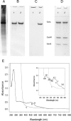
Analysis of CO dehydrogenase purified from O. carboxidovorans OM5 D::km. A, cell-free crude extract (15 μg) of O. carboxidovorans OM5 D::km grown with H2, CO2, CO, and air (as detailed in the legend to Fig. 3) was analyzed on native PAGE. B, native PAGE stained for protein with Coomassie Brilliant Blue of CO dehydrogenase purified from the mutant (lane 1) or from wild-type bacteria (lane 2). C, native PAGE stained for CO oxidizing activity with CO and 1-phenyl-2-(4-iodophenyl)-3-(4-nitrophenyl)-2H-tetrazolium chloride of CO dehydrogenase purified from the mutant (lane 1) or from wild-type bacteria (lane 2). D, denaturing PAGE stained for protein of CO dehydrogenase purified from the mutant (lane 1) or from wild-type bacteria (lane 2). Each lane contained 15 μg of CO dehydrogenase. CoxL, CoxM, and CoxS are the CO dehydrogenase subunits. E, UV-visible absorption spectra of apo-CO dehydrogenase: air-oxidized, trace a; reduced with pure CO for 30 min, trace b; reduced with 650 μm dithionite under N2 for 4 min, trace c. The inset shows the visible part of the spectra, including characteristic wavelengths (in nanometers). For experimental details refer to the “Experimental Procedures.”
FIGURE 5.
FIGURE 5.
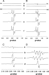
Analysis by EPR of iron-sulfur (A and B) and molybdenum (C and D) in CO dehydrogenase from mutant and wild type. Samples in 50 mm Tris-HCl, pH 8.2, contained 11.6 mg of protein/ml. Microwave frequency, modulation amplitude, and microwave power were 9.47 GHz, 1 millitesla, and 10 milliwatts, respectively. Spectra were recorded at 16 K (A), 50 K (B), and 120 K (C and D). A and B: traces a and b, air-oxidized; c and d, CO-reduced; e and f, reduced with 5 mm sodium dithionite; traces a, c, and e, CO dehydrogenase D::km; b, d, and f, wild-type CO dehydrogenase 23 units/mg. C, apo-CO dehydrogenase and wild-type (D). Traces a, air-oxidized; b, CO-reduced; c, reduced with 5 mm sodium dithionite; d, treated with 5 mm KCN for 24 h to remove cyanolysable sulfur and then reduced with 5 mm sodium dithionite.
FIGURE 6.
FIGURE 6.
Mo-K-edge EXAFS and corresponding Fourier transforms of CO dehydrogenase purified from O. carboxidovorans OM5 D::km. Experimental data are shown by black lines; calculated spectra are shown by red curves (A). Bond lengths are in Å (B).
FIGURE 7.
FIGURE 7.
Functional reconstruction of the non-functional [MoO3]-center in CO dehydrogenase D::km. Apo-CO dehydrogenase (2.4 mg/ml) was subjected to the following treatments: ○, sodium sulfide and sodium dithionite (5 mm each); •, 125 μm Cu+-thiourea first and then with sulfide plus dithionite; ▵, 125 μm Cu+-thiourea; and □, sodium sulfide and dithionite (5 mm each) followed by 125 μm Cu+-thiourea.
FIGURE 8.
FIGURE 8.
Recombinant CoxD of E. coli K38 pGP1-2/pETMW2 and subcellular localization of CoxD in O. carboxidovorans. A, formation of recombinant CoxD in E. coli K38 pGP1-2/pETMW2 was analyzed by SDS-PAGE of crude extracts (lanes 1 and 2). Bacteria grown for 210 min to A600 of 0.75 (lane 1; 20 μg of protein) were supplied with isopropyl-β-d-thiogalactopyranoside and grown for further 55 min at 30 °C. Then, T7 RNA polymerase was induced by raising the temperature to 42 °C for 90 min, and bacteria (A600 = 3.95) were analyzed for the formation of recombinant CoxD polypeptide (lane 2; 30 μg of protein). Cytoplasmic membranes of O. carboxidovorans (60 μg per lane) grown with H2, CO2, and CO were stained for protein on SDS-PAGE (lane 3) and analyzed for the presence of CoxD employing anti-CoxD IgG antibodies (lane 4). For details refer to the methods section. B, cell-free crude extracts of O. carboxidovorans were centrifuged in a sucrose density gradient (□) and analyzed for protein employing the Biuret-reaction (•), for CoxD by Western blotting (○), and for ATPase (▵) or GTPase (▴) activity by quantitation of inorganic phosphate.

Similar articles

Cited by

References

    1. Auling, G., Busse, J., Hahn, M., Hennecke, H., Kroppenstedt, R., Probst, A., and Stackebrandt, E. (1988) Syst. Appl. Microbiol. 10 264-272
    1. Meyer, O. O. (2005) in Bergey′s Manual of Systematic Bacteriology (Garrity, G. M., ed) pp. 468-471, vol. 2, Springer, New York
    1. Fuhrmann, S., Ferner, M., Jeffke, T., Henne, A., Gottschalk, G., and Meyer, O. (2003) Gene (Amst.) 322 67-75 - PubMed
    1. Paul, D., Bridges, S., Burgess, S. C., Dandass, Y., and Lawrence, M. L. (2008) J. Bacteriol. 190 5531-5532 - PMC - PubMed
    1. Santiago, B., Schübel, U., Egelseer, C., and Meyer, O. (1999) Gene (Amst.) 236 115-124 - PubMed

MeSH terms

LinkOut - more resources