. 2009 Apr;37(6):1886-96.
doi: 10.1093/nar/gkp036. Epub 2009 Feb 3.

A minimized rRNA-binding site for ribosomal protein S4 and its implications for 30S assembly

Affiliations

A minimized rRNA-binding site for ribosomal protein S4 and its implications for 30S assembly

Deepti L Bellur et al. Nucleic Acids Res. 2009 Apr.

Abstract

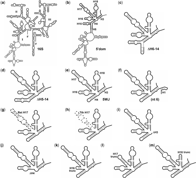
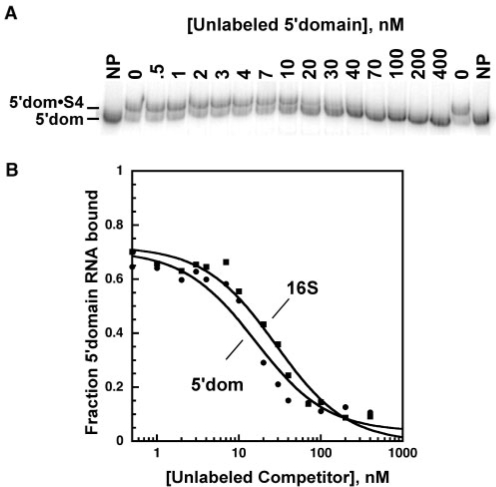
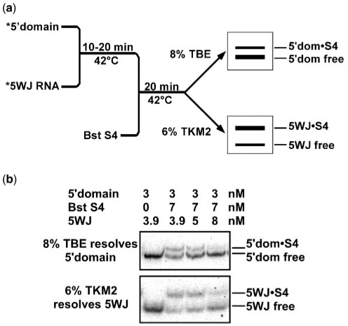
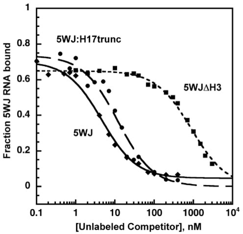
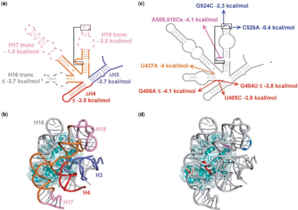
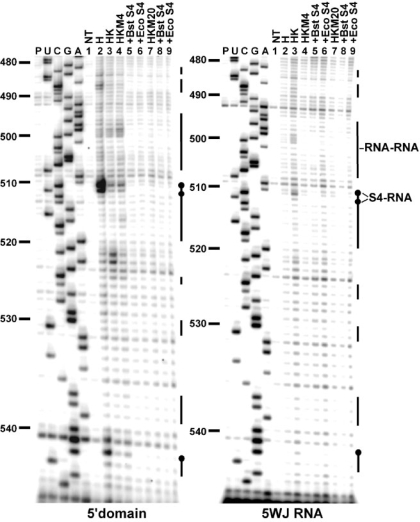
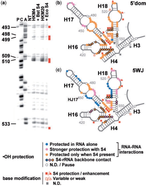
Primary ribosomal protein S4 is essential for 30S ribosome biogenesis in eubacteria, because it nucleates subunit assembly and helps coordinate assembly with the synthesis of its rRNA and protein components. S4 binds a five-helix junction (5WJ) that bridges the 5' and 3' ends of the 16S 5' domain. To delineate which nucleotides contribute to S4 recognition, sequential deletions of the 16S 5' domain were tested in competitive S4-binding assays based on electrophoretic mobility shifts. S4 binds the minimal 5WJ RNA containing just the five-helix junction as well or better than with affinity comparable to or better than the 5' domain or native 16S rRNA. Internal deletions and point mutations demonstrated that helices 3, 4, 16 and residues at the helix junctions are necessary for S4 binding, while the conserved helix 18 pseudoknot is dispensable. Hydroxyl radical footprinting and chemical base modification showed that S4 makes the same interactions with minimal rRNA substrates as with the native 16S rRNA, but the minimal substrates are more pre-organized for binding S4. Together, these results suggest that favorable interactions with S4 offset the energetic penalty for folding the 16S rRNA.


Figures

Figure 1.
Figure 1.
Escherichia coli 16S rRNA fragments tested for S4 binding. (a) 16S rRNA; (b) 5′domain; (c) RNA(▵H6-14); (d) RNA(▵H5-14); (e) 5WJ RNA; (f) 5WJ_nt6; (g) 5WJ:BstH17; (h) 5WJ:TthH17; (i) 5WJ▵H3; (j) 5WJ▵H4; (k) 5WJ:H16trunc; (l) 5WJ:H17trunc; and (m) 5WJ:H18trunc. Secondary structures of ▵H6-14 and 5WJ were confirmed by RNase T1 digestion (Figures S1 and S2).
Figure 2.
Figure 2.
Competitive binding of Bst S4 to the 16S 5′ domain RNA. (a) S4 binding measured by gel mobility shift. 0.5 nM 32P-labeled and 0–400 nM unlabeled 5′domain RNAs were incubated with 31.5 nM Bst S4 in HKM4 buffer at 42°C. Gel is 8% polyacrylamide in TBE. NP, 32P-5′domain RNA only. (b) The fraction of complexed 32P-labeled 5′domain RNA versus competitor RNA was fit to Equation (1). Filled circles, 5′ domain (Kd, 5′domain = 5.5 ± 2.6 nM); filled squares, 16S rRNA (Krel = 1.6 ± 0.8).
Figure 3.
Figure 3.
Dual-label competitive binding assay. (a) Scheme of the competitive binding experiments with two labeled RNAs. Complexes of the 5′ domain were separated on 8% polyacrylamide gels in TBE, while complexes of the 5WJ variants were separated on 6% polyacrylamide gels in TKM2, as described in ‘Material and Methods’ sections. (b) Distribution of S4 between 5′ domain and 5WJ RNAs. The counts in each complex and in free RNA were quantified and used to calculate Krel [Equation (2)]. For the 5WJ RNA, average Krel = 0.6 ± 0.1. The amount of labeled 5WJ complex decreases as unlabeled 5WJ RNA is added because its specific activity decreases. See Table 1 for further data.
Figure 4.
Figure 4.
Competitive binding of Bst S4 to 5WJ RNA. Representative plot of competition between 32P-labeled 5WJ RNA and unlabeled RNAs with 22.5 nM Bst S4 in HKM4 buffer. Diamonds, WT 5WJ (Kd = 0.72 ± 0.20 nM); circles, 5WJ:H17trunc (Krel = 4.1 ± 3.3); squares, 5WJΔH3 (Krel = 360 ± 130).
Figure 5.
Figure 5.
Sequence requirements for S4 binding. Energetic costs of deletions and point mutations in the 5WJ were obtained from competitive S4-binding assays against 32P-labeled 5WJ RNA at 42°C in HKM4 buffer. See Table S2 for further information. (a,b) Deletions in the 5WJ, and differences in dissociation free energy (ΔΔG), relative to the parental 5WJ RNA. (c,d) Base substitutions, as in (a). (b) and (d) Tertiary structure of 5WJ•S4 showing the deletions and mutations, colored as in (a) and (c), respectively, with S4 in cyan. Ribbons made with PDB 2AVY (41) using PyMOL (DeLano Scientific, http://www.pymol.org).
Figure 6.
Figure 6.
Hydroxyl radical footprinting on 5′ domain and 5WJ RNAs with S4. Representative footprinting gels covering nucleotides 480–544. The RNAs were probed under different buffer conditions at 42°C (lanes 4–6, HKM4; lanes 7–9, HKM20), with Bst (lanes 5 and 8) and Eco (lanes 6 and 9) S4. UCGA, dideoxy sequence ladders; P, reverse transcriptase pausing control; NT, untreated RNA. The vertical bars next to the lanes represent regions of protection from hydroxyl radical cleavage. Black bars, RNA–RNA interactions, black circles, S4–RNA interactions.
Figure 7.
Figure 7.
Structure probing of 5′domain and 5WJ RNAs with S4. (a) Representative sequencing gel showing DMS modification of 5WJ•S4 complex. The gel is labeled as in Figure 6, and symbols are as described in the key. (b,c) Summary of hydroxyl radical footprinting and chemical base modification on (b) 5′domain and (c) 5WJ RNA. Colored circles indicate backbone protections in HKM4. Colored circles with black outlines indicate riboses that are in direct contact with S4 [1j5e, (18); 2i2p, (42)]. Gray symbols, undetermined. Moderate-to-strong protection (squares) or enhanced base modification (triangles) is represented by closed red symbols. Open red symbols indicate Gs weakly or inconsistently modified by kethoxal.

Similar articles

Cited by

References

    1. Olsson MO, Isaksson LA. Analysis of rpsD mutations in Escherichia coli. III. Effects of rpsD mutations on expression of some ribosomal protein genes. Mol. Gen. Genet. 1979;169:271–278. - PubMed
    1. Yates JL, Arfsten AE, Nomura M. In vitro expression of Escherichia coli ribosomal protein genes: autogenous inhibition of translation. Proc. Natl Acad. Sci. USA. 1980;77:1837–1841. - PMC - PubMed
    1. Deckman IC, Draper DE. Specific interaction between ribosomal protein S4 and the alpha operon messenger RNA. Biochemistry. 1985;24:7860–7865. - PubMed
    1. Tang CK, Draper DE. Evidence for allosteric coupling between the ribosome and repressor binding sites of a translationally regulated mRNA. Biochemistry. 1990;29:4434–4439. - PubMed
    1. Spedding G, Draper DE. Allosteric mechanism for translational repression in the Escherichia coli alpha operon. Proc. Natl Acad. Sci. USA. 1993;90:4399–4403. - PMC - PubMed

Publication types

MeSH terms