. 2009 Apr 9;113(15):3577-84.
doi: 10.1182/blood-2008-09-178913. Epub 2009 Feb 3.

Loss of pleckstrin defines a novel pathway for PKC-mediated exocytosis

Affiliations

Loss of pleckstrin defines a novel pathway for PKC-mediated exocytosis

Lurong Lian et al. Blood. .

Abstract

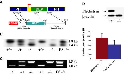
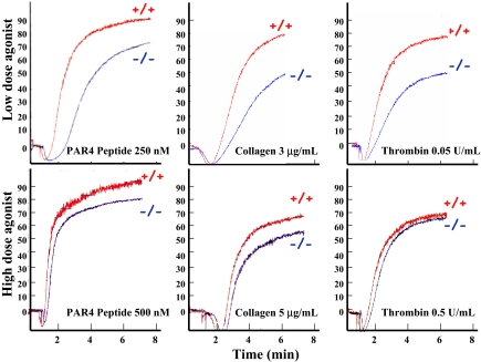
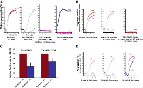
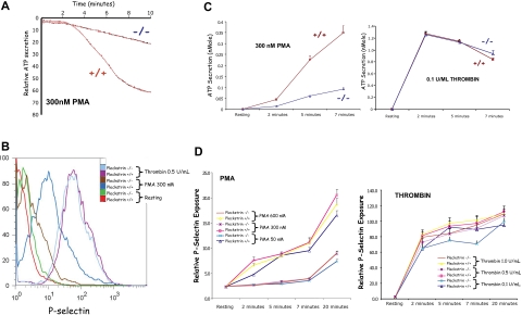
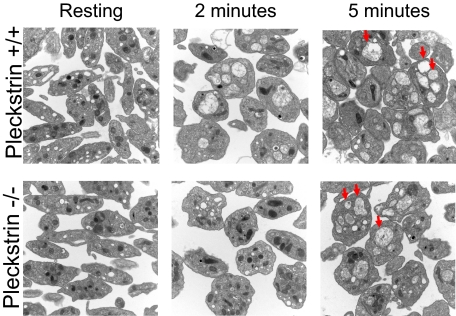
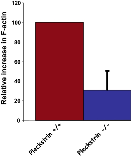
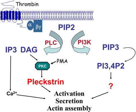
Pleckstrin, the platelet and leukocyte C kinase substrate, is a prominent substrate of PKC in platelets, monocytes, macrophages, lymphocytes, and granulocytes. Pleckstrin accounts for 1% of the total protein in these cells, but it is best known for containing the 2 prototypic Pleckstrin homology, or PH, domains. Overexpressed pleckstrin can affect polyphosphoinositide second messenger-based signaling events; however, its true in vivo role has been unknown. Here, we describe mice containing a null mutation within the pleckstrin gene. Platelets lacking pleckstrin exhibit a marked defect in exocytosis of delta and alpha granules, alphaIIbbeta3 activation, actin assembly, and aggregation after exposure to the PKC stimulant, PMA. Pleckstrin-null platelets aggregate normally in response to thrombin, but they fail to aggregate in response to thrombin in the presence of PI3K inhibitors, suggesting that a PI3K-dependent signaling pathway compensates for the loss of pleckstrin. Although pleckstrin-null platelets merged their granules in response to stimulation of PKC, they failed to empty their contents into the open canalicular system. This might be attributable to impaired actin assembly present in cells lacking pleckstrin. These data show that pleckstrin regulates the fusion of granules to the cell membrane and is an essential component of PKC-mediated exocytosis.


Figures

Figure 1
Figure 1
Pleckstrin targeting. (A) Recombination into the pleckstrin gene results in the deletion of the nucleotides corresponding to amino acids 76 through 128 encoded within exon 3. This removes the distal portion of a β sheet, and the critical α helix from the first PH domain. (B) Southern blotting of mouse genomic DNA. Southern blotting shows a 2.8-kb wild-type band, and a 2.4-kb targeted band. (C) PCR of genomic DNA with a sense primer homologous to a region 5′ of the targeted region and antisense primers homologous to either HPRT or exon 3 of the pleckstrin gene. (D) Antipleckstrin immunoblot of platelet lysates showing complete loss of pleckstrin protein in mice homozygous for the recombinant gene. (E) The mean and SD of platelet counts derived from 8 pleckstrin-null mice and 8 wild-type littermates are shown.
Figure 2
Figure 2
Pleckstrin-null platelets have a PKC-mediated aggregation defect. Washed murine platelets lacking pleckstrin were analyzed after agonist stimulation in a Lumi-Aggregometer. The y-axis shows the relative aggregation, and the x-axis shows time in minutes. Platelets lacking pleckstrin have nearly normal aggregation in response to high doses of a peptide agonist of PAR4 (the dominant murine thrombin receptor), collagen, and thrombin (bottom panels). In contrast, lower doses of these agonists show that pleckstrin-null platelets have a mild aggregation defect (top). Results are representative of 6 experiments.
Figure 3
Figure 3
PKC-mediated aggregation is absent in pleckstrin-null platelets. (A) In contrast to their response to thrombin, washed pleckstrin-null platelets completely fail to aggregate in response to all doses of the PKC stimulant, PMA (25-600 nM). The third tracing from the left shows that the effect of PMA on platelet aggregation is completely ablated by the PKC inhibitor, GF109203x. The dose response was derived from 3 experiments. (B) The presence of a PI3K inhibitor completely eliminates the ability of pleckstrin-null platelets to aggregate in response to the peptide agonist of PAR4 (250 μM). Inhibition of wild-type platelets with both a PI3K inhibitor (LY294002) and PKC inhibitor (GF109203x) resulted in a total loss of aggregation in response to 250 μM of the PAR4 peptide agonist. The results are representative of 3 experiments. (C) Loss of pleckstrin also impairs the binding of Jon/A, an antibody that only recognizes the activated form of αIIbβ3. The mean and SD are derived from 6 experiments with PMA (P < .001) and 10 experiments with thrombin (P < .001). (D) The addition of exogenous fibrinogen reverts the aggregation defect of washed platelets lacking pleckstrin in response to 300 nM PMA. These results are representative of 3 experiments.
Figure 4
Figure 4
PKC-mediated exocytosis is impaired in platelets lacking pleckstrin. PMA-induced exocytosis was analyzed biochemically (A,C), as well as by flow cytometry (B,D). The effect of the pleckstrin loss on function mutation on the δ (dense) granule secretion was analyzed in a Lumi-Aggregometer (Chrono-Log). (A) A representative tracing is shown, and pooled results showing the mean and SD derived from 5 experiments for PMA and 4 experiments with thrombin are plotted (C). Platelets lacking pleckstrin had a consistent deficit in dense granule section (P < .01 for all time points). The flow histogram (B) shows the relative surface exposure of P-selectin as a marker of α granule fusion with the cell membrane. The pooled analysis derived from 4 experiments is shown (D). For all doses and time points, paired analysis showed a P < .05.
Figure 5
Figure 5
Loss of pleckstrin allows granule-to-granule fusion. Morphologies of platelet granules were analyzed in resting platelets and in platelets exposed to PMA for 2 or 5 minutes. Platelets derived from wild-type or pleckstrin-null platelets appeared identical under basal conditions. Wild-type platelets coalesced their granules more rapidly than platelets lacking pleckstrin (compare images in middle column.) However, wild-type and pleckstrin-null platelets appeared identical after 5 minutes of stimulation, with both genotypes of platelets having numerous merged granules (several examples indicated by arrowheads.) This shows that loss of pleckstrin impairs the efficiency of granule-to-granule fusion but does not prevent it. Shown are 20 000× magnifications captured with an FEI Tecnai T12 electron microscope operated at 80-kV accelerating voltage.
Figure 6
Figure 6
Loss of pleckstrin impairs actin assembly. After stimulation of washed murine platelets with 300 nM PMA, platelets were fixed, permeabilized, and stained with fluorescent phalloidin. Flow cytometry was used to quantitate phalloidin binding in 100 000 cells, and analysis was performed using FlowJo software. Shown is the mean plus or minus SEM for 5 experiments (P < .015).
Figure 7
Figure 7
Model of signaling by pleckstrin leading to platelet activation. Stimulation of an agonist receptor leads to the activation of PI3K and PLC. These 2 enzymes participate in alternative signaling pathways, leading to platelet aggregation. Activation of PLC leads to the production of DAG, PKC activation, and pleckstrin phosphorylation. Once phosphorylated, pleckstrin binds to the cell membrane and contributes to integrin activation, actin assembly, and exocytosis. An alternative pathway to platelet activation involving PI3K is pleckstrin-independent.

References

    1. Haslam RJ, Lynham JA, Fox JEB. Effects of collagen, ionophore A23187 and prostaglandin E1 on the phosphorylation of specific proteins in blood platelets. Biochem J. 1979;178:397–406. - PMC - PubMed
    1. Imaoka T, Lynham JA, Haslam RJ. Purification and characterization of the 47,000-dalton protein phosphorylated during degranulation of human platelets. J Biol Chem. 1983;258:11404–11414. - PubMed
    1. Connolly TM, Lawing WJJ, Majerus PW. Protein kinase C phosphorylates human platelet inositol trisphosphate 5′-phosphomonoesterase, increasing the phosphatase activity. Cell. 1986;46:951–958. - PubMed
    1. Tyers M, Haslam RJ, Rachubinski RA, Harley CB. Molecular analysis of pleckstrin: the major protein kinase C substrate of platelets. J Cell Biochem. 1989;40:133–145. - PubMed
    1. Tyers M, Rachubinski RA, Stewart MI, et al. Molecular cloning and expression of the major protein kinase C substrate of platelets. Nature. 1988;333:470–473. - PubMed

Publication types