. 2009 Apr;296(4):E888-97.
doi: 10.1152/ajpendo.90946.2008. Epub 2009 Feb 3.

Diurnal expression of functional and clock-related genes throughout the rat HPA axis: system-wide shifts in response to a restricted feeding schedule

Affiliations

Diurnal expression of functional and clock-related genes throughout the rat HPA axis: system-wide shifts in response to a restricted feeding schedule

Milena Girotti et al. Am J Physiol Endocrinol Metab. 2009 Apr.

Abstract

The diurnal rhythm of glucocorticoid secretion depends on the suprachiasmatic (SCN) and dorsomedial (putative food-entrainable oscillator; FEO) nuclei of the hypothalamus, two brain regions critical for coordination of physiological responses to photoperiod and feeding cues, respectively. In both cases, time keeping relies upon diurnal oscillations in clock gene (per1, per2, and bmal) expression. Glucocorticoids may play a key role in synchronization of the rest of the body to photoperiod and food availability. Thus glucocorticoid secretion may be both a target and an important effector of SCN and FEO output. Remarkably little, however, is known about the functional diurnal rhythms of the individual components of the hypothalamic-pituitary-adrenal (HPA) axis. We examined the 24-h pattern of hormonal secretion (ACTH and corticosterone), functional gene expression (c-fos, crh, pomc, star), and clock gene expression (per1, per2 and bmal) in each compartment of the HPA axis under a 12:12-h light-dark cycle and compared with relevant SCN gene expression. We found that each anatomic component of the HPA axis has a unique circadian signature of functional and clock gene expression. We then tested the susceptibility of these measures to nonphotic entrainment cues by restricting food availability to only a portion of the light phase of a 12:12-h light-dark cycle. Restricted feeding is a strong zeitgeber that can dramatically alter functional and clock gene expression at all levels of the HPA axis, despite ongoing photoperiod cues and only minor changes in SCN clock gene expression. Thus the HPA axis may be an important mediator of the body entrainment to the FEO.


Figures
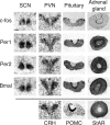
Fig. 1.
Fig. 1.
Representative autoradiogram images of functional and clock gene expression measured by in situ hybridization. Images were selected from cases corresponding to the time of maximal expression of the hormone producing functional genes, corticotropin-releasing hormone gene (crh) heteronuclear RNA (hnRNA), proopiomelanocortin gene (pomc) hnRNA, and steroidogenic acute regulatory protein gene (star) mRNA, in the paraventricular nucleus of the hypothalamus (PVN), pituitary, and adrenal components of the hypothalamus-pituitary-adrenal (HPA) axis, respectively, and of an immediate early gene (c-fos mRNA) and clock genes [period gene (per) 1 mRNA, per2 mRNA, and brain and muscle aryl hydrocarbon receptor nuclear translocator-like gene (bmal) mRNA] in the suprachiasmatic nucleus (SCN), PVN, pituitary, and adrenal gland.
Fig. 2.
Fig. 2.
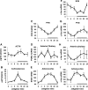
Best-fit circadian rhythm parameters and statistical analyses for the hormone and gene expression measures in experiment 1. The phase angle (x0) with 95% confidence interval (CI) for each hormone and gene expression measure of experiment 1 (also see Fig. 3) were estimated and the values graphically presented (A) and tabulated (B). In B, the estimates of the measured variable's maximum (max) and minimum (min) and the F and P values of the circadian rhythm best fit (24-h sinusoid function) are also presented. The x0, CI, max, and min are expressed in zeitgeber time (ZT). +Cases of per1 or per2 gene expression that have phase angles that occur 12 h in antiphase with bmal gene expression in the same tissue. In A, the dark bar above the x-axis represents the period of lights off. AP and Ant Pit, anterior pituitary; Adr and Adrcort, adrenocortex.
Fig. 3.
Fig. 3.
HPA axis hormone secretion and functional gene expression change over 24 h. Adrenocorticotropic hormone (ACTH; A) and corticosterone (B) plasma levels were measured over a 24-h period (n = 6). The crh (C), pomc (D) hnRNA, star (E), and c-fos (F-I) mRNA levels in tissue from the same rats were determined by in situ hybridization and are expressed as percent of ZT0. The ZT0 measure has been plotted two times (ZT0 and ZT24) for graphical visualization purposes. The dark bar above the x-axis represents the period of lights off.
Fig. 4.
Fig. 4.
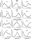
Clock gene expression has specific temporal patterns in the SCN and in the individual components of the HPA axis. The levels of per1, per2, and bmal mRNAs were determined by in situ hybridization in the SCN (A-C, respectively), PVN (D-F), anterior pituitary (G-I), and adrenocortex (J-L) over 24 h (n = 6) and are expressed as percent of ZT0. The ZT0 measure has been plotted two times (ZT0 and ZT24) for graphical visualization purposes. The dark bar above the x-axis represents the period of lights off.
Fig. 5.
Fig. 5.
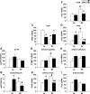
Restricted feeding (RF) affects the diurnal rhythms of corticosterone and functional gene expression in the HPA axis. Rats were either fed ad libitum (AL) or fed for 3 h between ZT3 and ZT6 (RF). ACTH (A) and corticosterone (B) levels were measured in the plasma, and the crh (C) and pomc (D) hnRNA and star (E) and c-fos (F-I) mRNA levels were determined at ZT2 (A.M.) and ZT11 (P.M.) in the indicated tissues (n = 6). Gene expression levels are represented as percent of the morning ad libitum levels (AL-A.M.). *Significant difference between A.M. and P.M. times within the same feeding condition. #Significant difference between the same time of day in the different feeding conditions, as established by Fisher's least-significant difference (LSD) test (P < 0.05).
Fig. 6.
Fig. 6.
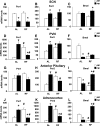
RF substantially affects the diurnal expression of clock genes in the HPA axis, with limited effect in the SCN. Rats (n = 6) were either fed ad libitum or fed for 3 h between ZT3 and ZT6 (RF). The levels of per1, per2, and bmal mRNAs were determined in the SCN (A-C, respectively), PVN (D-F), anterior pituitary (G-I), and adrenocortex (J-L) at ZT2 (A.M.) and ZT11 (P.M.). Gene expression levels are represented as percent of the morning ad libitum levels (AL-A.M.). *Significant difference between A.M. and P.M. times within the same feeding condition. #Significant difference between the same time of day in the different feeding conditions, as established by Fisher's LSD test (P < 0.05).

References

    1. Abe M, Herzog ED, Yamazaki S, Straume M, Tei H, Sakaki Y, Menaker M, Block GD. Circadian rhythms in isolated brain regions. J Neurosci 22: 350–356, 2002. - PMC - PubMed
    1. Amir S, Lamont EW, Robinson B, Stewart J. A circadian rhythm in the expression of PERIOD2 protein reveals a novel SCN-controlled oscillator in the oval nucleus of the bed nucleus of the stria terminalis. J Neurosci 24: 781–790, 2004. - PMC - PubMed
    1. Angeles-Castellanos M, Aguilar-Roblero R, Escobar C. c-Fos expression in hypothalamic nuclei of food-entrained rats. Am J Physiol Regul Integr Comp Physiol 286: R158–R165, 2004. - PubMed
    1. Asai M, Yoshinobu Y, Kaneko S, Mori A, Nikaido T, Moriya T, Akiyama M, Shibata S. Circadian profile of Per gene mRNA expression in the suprachiasmatic nucleus, paraventricular nucleus, and pineal body of aged rats. J Neurosci Res 66: 1133–1139, 2001. - PubMed
    1. Balsalobre A, Brown SA, Marcacci L, Tronche F, Kellendonk C, Reichardt HM, Schutz G, Schibler U. Resetting of circadian time in peripheral tissues by glucocorticoid signaling. Science 289: 2344–2347, 2000. - PubMed

Publication types

MeSH terms

LinkOut - more resources