. 2009 Feb 15;69(4):1375-82.
doi: 10.1158/0008-5472.CAN-08-2896. Epub 2009 Feb 3.

Identification of a novel pathway that selectively modulates apoptosis of breast cancer cells

Affiliations

Identification of a novel pathway that selectively modulates apoptosis of breast cancer cells

Alexander A Tinnikov et al. Cancer Res. .

Abstract

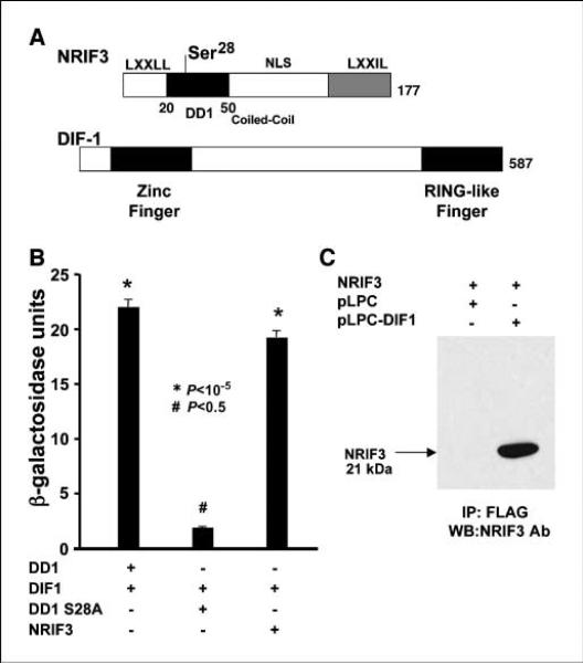
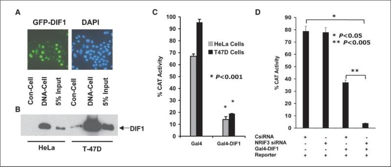
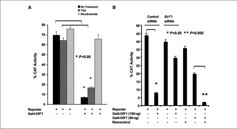
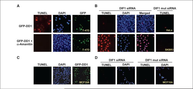
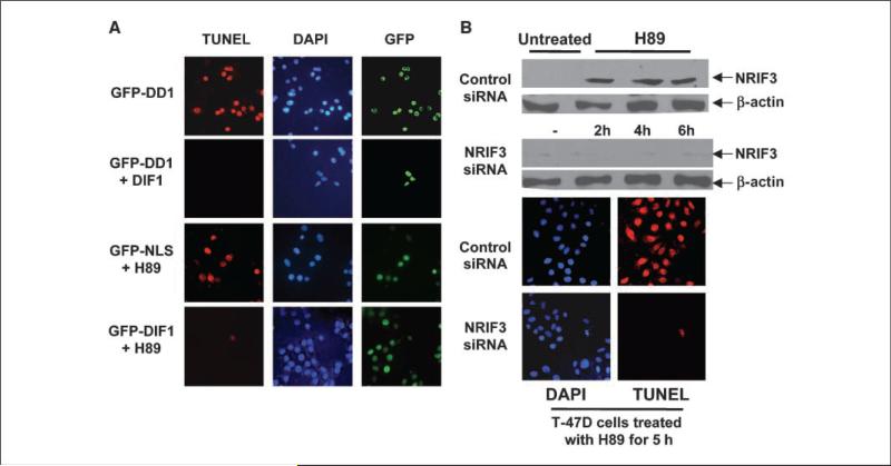
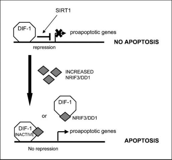
Expression of the nuclear receptor interacting factor 3 (NRIF3) coregulator in a wide variety of breast cancer cells selectively leads to rapid caspase-2-dependent apoptotic cell death. A novel death domain (DD1) was mapped to a 30-amino acid region of NRIF3. Because the cytotoxicity of NRIF3 and DD1 seems to be cell type-specific, these studies suggest that breast cancer cells contain a novel "death switch" that can be specifically modulated by NRIF3 or DD1. Using an MCF-7 cell cDNA library in a yeast two-hybrid screen, we cloned a factor that mediates apoptosis by DD1 and refer to this factor as DD1-interacting factor-1 (DIF-1). DIF-1 is a transcriptional repressor that mediates its effect through SirT1, and this repression is attenuated by the binding of NRIF3/DD1. DIF-1 expression rescues breast cancer cells from NRIF3/DD1-induced apoptosis. Small interfering RNA (siRNA) knockdown of DIF-1 selectively leads to apoptosis of breast cancer cells, further suggesting that DIF-1 plays a key role in NRIF3/DD1-mediated apoptosis. A protein kinase A inhibitor (H89) also elicits apoptosis of breast cancer cells but not of the other cell types examined, and DIF-1 also protects these cells from H89-mediated apoptosis. In addition, H89 incubation results in a rapid increase in NRIF3 levels and siRNA knockdown of NRIF3 protects breast cancer cells from H89-mediated apoptosis. Our results indicate that DIF-1 plays a key role in breast cancer cell survival and further characterizing this pathway may provide important insights into developing novel therapies to selectively target breast cancer cells for apoptosis.


Figures

Figure 1
Figure 1
Structure, domains, and interaction of NRIF3 and DIF-1. A, the domain structure of NRIF3 has been described previously (8). The DD1 region sufficient to mediate apoptosis (amino acids 20–50) is shown along with Ser28, which is essential to mediate apoptosis. DIF-1 contains an NH2 terminal zinc finger and a COOH terminal RING-like finger. DD1 interacts with DIF-1 through the RING-like finger. B and C, NRIF3/DD1 interacts with DIF-1 in yeast and mammalian cells. B, LexA-NRIF3, LexA-DD1, and LexA-DD1(S28A) were expressed in yeast along with a LexA-driven LacZ reporter (pSH18-34) and pJG4-5 conditionally expressing DIF-1 with a B42 activation domain. Shown are LacZ results where B42-DIF-1 was conditionally expressed by galactose. LexA-NRIF3 and LexA-DD1 interact with DIF-1, whereas LexA-DD1(S28A) exhibited little or no interaction. B, HeLa cells were transfected with pLPC-DIF-1 or the control pLPC vector along with pcDNA3.1 expressing NRIF3. Twenty-four hours later, the cells were lysed and incubated overnight with FLAG-antibody (M2) agarose beads. The protein bound to the beads was analyzed by Western blotting with NRIF3 antibody.
Figure 2
Figure 2
DIF-1 is a nuclear protein that binds to DNA-cellulose and mediates transcriptional repression. A, GFP-DIF-1 expressed in T-47D cells localizes to the nucleus. B, HeLa and T-47D cells expressing FLAG-HA tagged DIF-1 were lysed in 0.3 mol/L KCl buffer. The supernatants were bound to FLAG antibody beads and then eluted with 3× FLAG peptide. Samples were incubated with DNA-cellulose (DNA-Cell) or with cellulose control (Con-Cell), and after washing, the cellulose beads were analyzed for DIF-1 by Western blotting with FLAG antibody. The input represents 5% of the sample before the binding assay. C, HeLa or T-47D cells were transfected with 100 ng of the pG5-SV-BCAT reporter along with 200 ng of vector expressing Gal4-DIF-1 or 150 ng (equal molar amount of plasmid) expressing only the Gal4 DBD. CAT activity was analyzed 30 h later. Gal4-DIF-1 represses the Gal4-CAT reporter in both cell types. D, HeLa cells were transfected as indicated with an NRIF3 siRNA (25 nmol/L) or a control-siRNA (25 nmol/L). Thirty hours later, the cells were transfected with 100 ng of pG5-SV-BCAT alone and with 50 ng of the Gal4-DIF-1 vector. CAT activity was determined 30 h later.
Figure 3
Figure 3
Repression by DIF-1 is mediated by SirT1. A, T-47D cells were transfected with 100 ng of pG5-SV-BCAT and 200 ng of vector expressing Gal4-DIF-1. One hour before transfection, cells received either TSA (150 ng/mL), nicotinamide (50 nmol/L), or no treatment. Thirty hours later, the cells were harvested for CAT activity. B, T-47D cells were transfected with a SirT1 siRNA (25 nmol/L) or a control-siRNA (25 nmol/L). Before introduction of the siRNAs, cells were incubated with 20 μmol/L zVDVAD-fmk, a caspase-2 inhibitor, to prevent apoptosis that might occur as a result of knockdown of SirT1. Thirty hours after introduction of the siRNAs, the cells were transfected with 100 ng of the pG5-SV-BCAT reporter alone and with 150 ng of the Gal4-DIF-1 expression vector. Another set of cells did not receive siRNA but received 1 μmol/L resveratrol before transfection with 50 ng of the Gal4-DIF-1 vector. Thirty hours after transfection, the cells were analyzed for CAT activity.
Figure 4
Figure 4
Effect of α-amanitin on DD1-mediated apoptosis and knockdown of DIF-1 by siRNA leads to apoptosis of breast cancer cells. A, T-47D cells were incubated with or without α-amanitin (2.5 μmol/L) for 3 h and then transfected with vector (100 ng) to express GFP-DD1. Twenty hours later, the cells were analyzed for GFP-DD1 expression and apoptosis by TUNEL assay. Shown are representative fields. No apoptosis was identified in cells preincubated with α-amanitin and the levels of GFPDD1 were similar under both conditions. B, siRNA (25 nmol/L) was used to knockdown DIF-1 expression in five different breast cancer cell lines (SKBR3, MCF-7, T-47D, MDA-435, and MDA-231) or cells of other origin (U2OS, human osteosarcoma; 293, human kidney epithelium; UOK-145, kidney carcinoma; HepG2, human hepatoma; HeLa, human cervical epithelium). A control siRNA (25 nmol/L) contained four base changes (mut siRNA). Thirty hours later, cells were examined for apoptosis by TUNEL assay. All the breast cancer cell lines exhibited apoptosis, whereas the other cells did not. Shown are representative results from the study with SKBR3 breast cancer cells and HeLa cells. Supplementary Fig. S1 shows results with the other cell lines. Similar results were found with four different siRNAs that targeted DIF-1 mRNA. C, expression of DD1 (GFP-DD1) does not lead to apoptosis of MCF10A cells, a cell line derived from normal breast epithelial cells. D, DIF-1 siRNA does not lead to apoptosis of MCF10A cells.
Figure 5
Figure 5
DIF-1 rescues breast cancer cells from DD1-mediated and H89-mediated apoptosis. A, T-47D cells were transfected with 100 ng pLPC-DIF-1. Other cells were transfected with control pLPC vector. Twenty-four hours later, the cells were transfected with 50 ng of GFP-DD1 vector. Five hours later, cells were analyzed for apoptosis by TUNEL assay. Shown are represented fields. Expression of DIF-1 decreased DD1-mediated apoptosis by over 90%. For the H89 study, T-47D cells were transfected with GFP-DIF-1 vector (100 ng) or with a vector expressing GFP containing a nuclear localization signal (GFP-NLS; 75 ng). Twenty-four hours later, the cells received 200 nmol/L of H89, a PKA inhibitor. Five hours later, the cells were examined for apoptosis by TUNEL assay. Shown are representative fields. Expression of DIF-1 resulted in a >90% reduction in apoptosis mediated by H89. B, H89 mediates apoptosis in breast cancer cells through NRIF3. T-47D cells were treated with a NRIF3 siRNA (25 nmol/L) or with a control siRNA (25 nmol/L). Thirty hours later, the cells received 200 nmol/L H89. Cells were harvested for NRIF3 expression by Western blotting 2, 4, and 6 h after addition of H89. Cells were also examined for apoptosis by TUNEL assay 5 h after H89 incubation. H89 incubation results in a rapid increase in NRIF3. NRIF3 siRNA blocked the rapid H89-mediated increase in NRIF3 expression, as well as apoptosis. Supplementary Fig. S5 indicates that this rapid increase in NRIF3 occurs at the mRNA level.
Figure 6
Figure 6
Mechanism by which NRIF3/DD1 and DIF-1 regulate apoptosis of breast cancer cells. The figure depicts a model that is consistent with our findings that DIF-1 is a transcriptional repressor that can bind DNA whose activity is reversed by NRIF3/DD1 and that preincubation with α-amanitin blocks DD1-mediated apoptosis of breast cancer cells. Our findings suggest that one or more proapoptotic genes bind to and are repressed by DIF-1 in breast cancer cells. Expression of NRIF3/DD1 binds DIF-1 and mediates a conformational change that reverses its ability to act as a repressor. We do not know whether the NRIF3/DIF-1 complex remains bound to the proapoptotic gene(s) or whether the binding of NRIF3/DD1 to DIF-1 results in a dissociation of DIF-1 from the proapoptotic gene. By either mechanism, reversal of repression of one or more proapoptotic genes leads to gene expression and the initiation of apoptosis.

References

    1. Hengartner MO. The biochemistry of apoptosis. Nature. 2000;407:770–6. - PubMed
    1. Lahm A, Paradisi A, Green DR, Melino G. Death fold domain interaction in apoptosis. Cell Death Differ. 2003;10:10–2. - PubMed
    1. Afonja A, Raaka BM, Huang A, et al. RAR agonists stimulate SOX9 gene expression in breast cancer cell lines: evidence for a role in retinoid-mediated growth inhibition. Oncogene. 2002;21:7850–60. - PubMed
    1. Afonja O, Juste D, Das S, Matsuhashi S, Samuels HH. Induction of PDCD4 tumor suppressor gene expression by RAR agonists, antiestrogen and HER-2/neu antagonist in breast cancer cells. Evidence for a role in apoptosis. Oncogene. 2004;23:8135–45. - PubMed
    1. Li D, Das S, Yamada T, Samuels HH. The NRIF3 family of transcriptional coregulators induces rapid and profound apoptosis in breast cancer cells. Mol Cell Biol. 2004;24:3838–48. - PMC - PubMed