. 2009 Feb 15;69(4):1416-28.
doi: 10.1158/0008-5472.CAN-08-0857. Epub 2009 Feb 3.

Trastuzumab reverses letrozole resistance and amplifies the sensitivity of breast cancer cells to estrogen

Affiliations

Trastuzumab reverses letrozole resistance and amplifies the sensitivity of breast cancer cells to estrogen

Gauri Sabnis et al. Cancer Res. .

Abstract

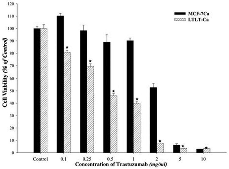
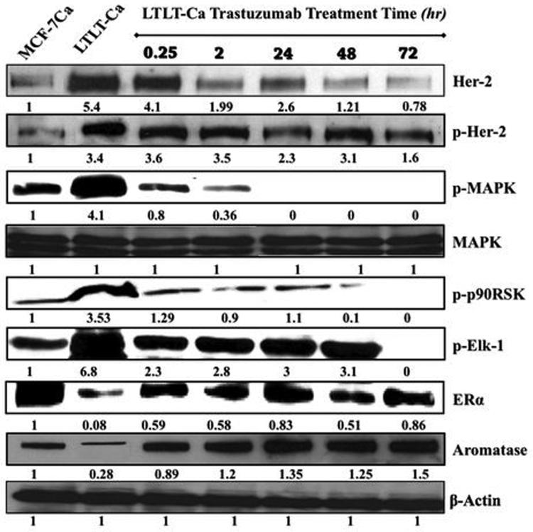
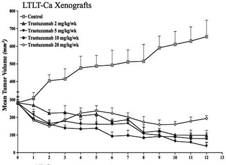
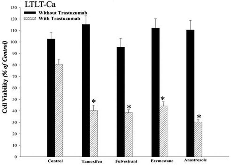
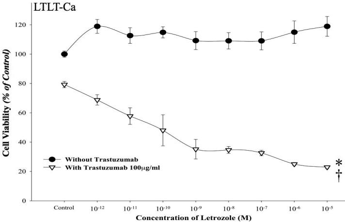
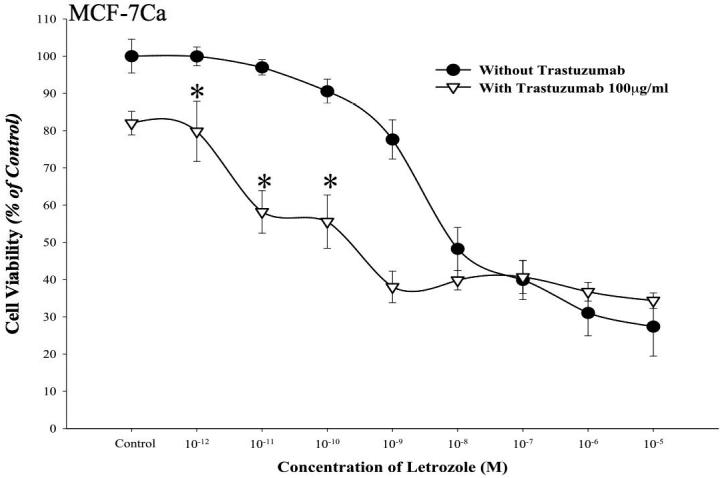
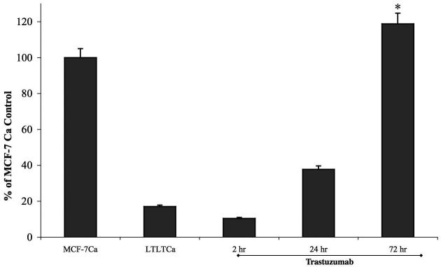
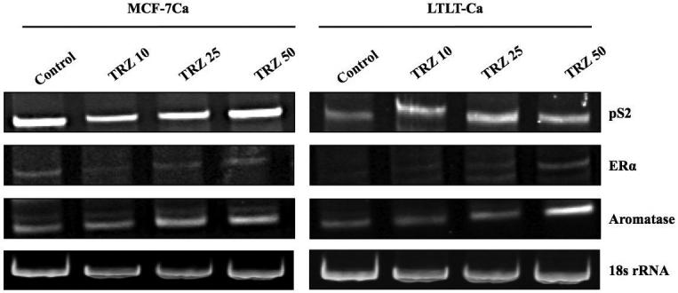
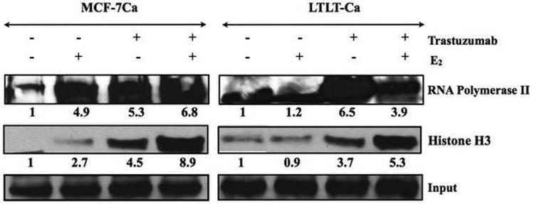
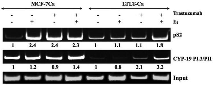
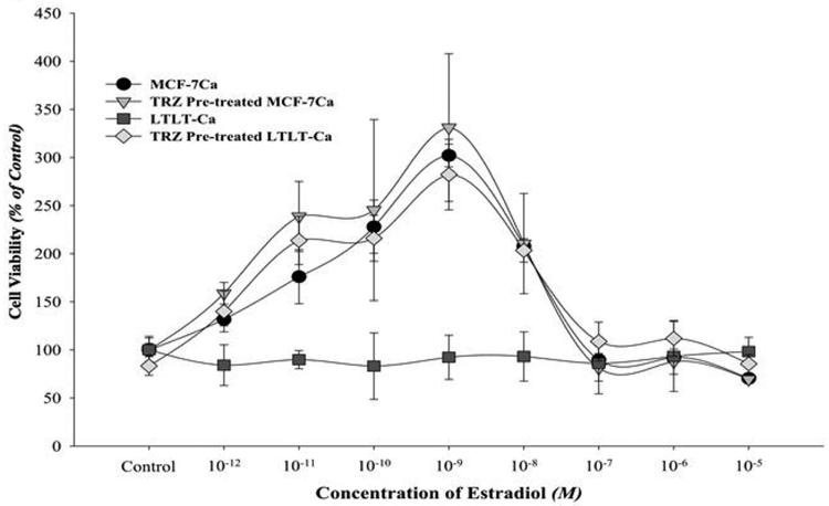
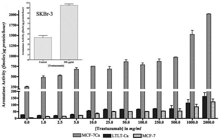
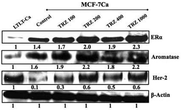
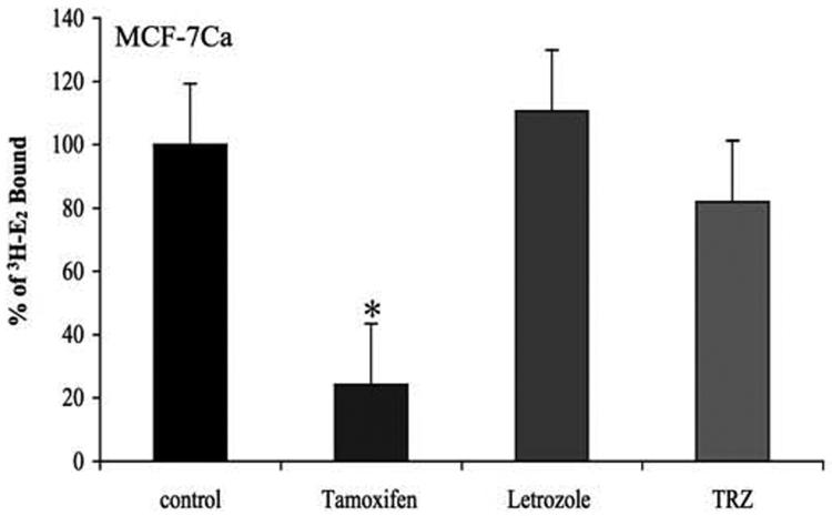
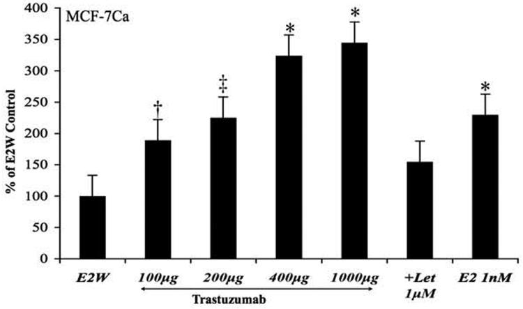
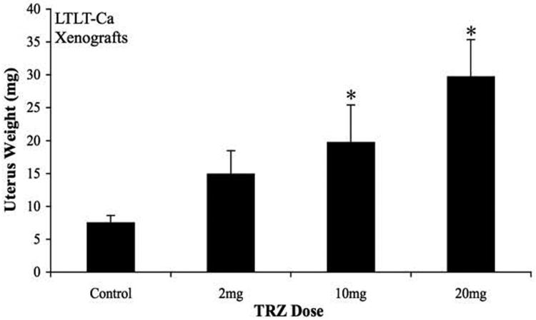
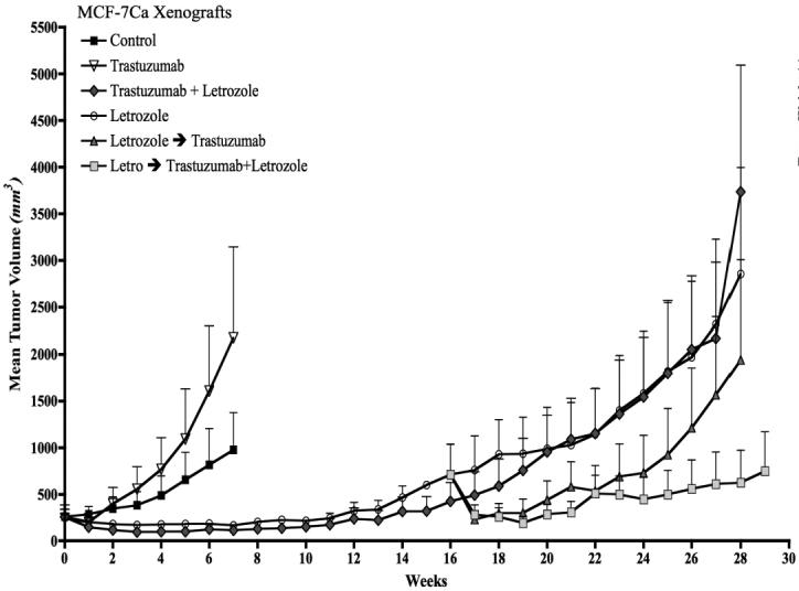
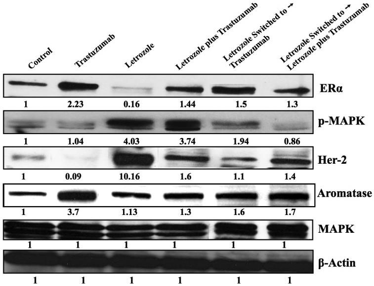
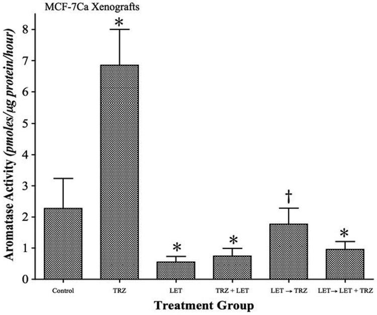
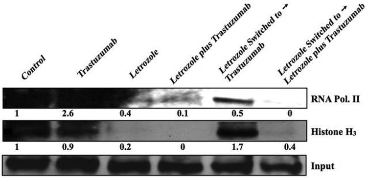
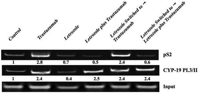
In this study, we investigated adaptive mechanisms associated with aromatase inhibitor (AI) resistance in breast cancer cells and show that sensitivity to AIs can be extended through dual inhibition of estrogen receptor (ER) and human epidermal receptor-2 (Her-2) signaling. We used human ER-positive breast cancer cells stably transfected with the aromatase gene (MCF-7Ca). These cells grow as tumors in nude mice and are inhibited by AIs. Despite continued treatment, tumors eventually become insensitive to AI letrozole. The cells isolated from these long-term letrozole-treated tumors (LTLT-Ca) were found to have decreased ERalpha levels. Our results suggest that LTLT-Ca cells survive estrogen deprivation by activation of Her-2/mitogen-activated protein kinase (MAPK) pathway. Here, we show that trastuzumab (antibody against Her-2; IC(50) = 0.4 mg/mL) was very effective in restoring the ERalpha levels and sensitivity of LTLT-Ca cells to endocrine therapy by down-regulation of Her-2/MAPK pathway and up-regulation of ERalpha. In contrast, trastuzumab was ineffective in the parental hormone-responsive MCF-7Ca cells (IC(50) = 4.28 mg/mL) and xenografts. By blocking Her-2, trastuzumab also up-regulates ERalpha and aromatase expression and hypersensitized MCF-7Ca cells to E(2). We show that trastuzumab is beneficial in hormone-refractory cells and xenografts by restoring ER, implicating Her-2 as a negative regulator of ERalpha. In xenograft studies, the combination of trastuzumab plus letrozole is equally effective in inhibiting growth of MCF-7Ca tumors as letrozole alone. However, on the acquisition of resistance and increased Her-2 expression, the combination of letrozole plus trastuzumab provided superior benefit over letrozole or trastuzumab alone.


Figures

Figure 1A
Figure 1A. Effect of trastuzumab on proliferation of MCF-7Ca and LTLT-Ca cells in vitro
Viability of cells was measured by MTT assay after 6 day treatment with trastuzumab as described in Materials and Methods. The treatment with trastuzumab is significantly more effective in reducing cell viability of LTLT-Ca cells compared to MCF-7Ca cells at all doses. At each dose of trastuzumab tested (0.1mg/ml through 10mg/ml) in LTLT-Ca cells growth was inhibited significantly better (*p-value <0.0001) than in MCF-7Ca cells.
Figure 1B
Figure 1B. Effect of trastuzumab treatment at various time points on the expression of protein in the Her-2/MAPK pathway, ERα and aromatase in LTLT-Ca cells
Expression of proteins was examined using western imunoblotting as described in materials and methods. Blot shows Her-2, p-Her-2 at 185 kDa, p-MAPK, MAPK at 42-44 kDa, phospho-p90RSK at 90 kDa, phospho-Elk-1 at 41 kDa, , ERα at 66 kDa, aromatase at 55 kDa and β-actin at 45 kDa. The blots show a single representative of three independent experiments.
Figure 1C
Figure 1C. Effect of trastuzumab treatment at various doses on the growth of LTLT-Ca xenografts
LTLT-Ca xenografts were grown in female OVX nude mice as described in Materials and Methods. The mice in the control and letrozole treated group exhibited similar rate of tumor growth. The difference in the exponential parameter governing growth was -0.027 (p = 0.71) over first eight weeks and 0.001 (p value = 0.97) over 19 weeks. Four doses of trastuzumab were tested. All of the tested doses caused a marked regression of LTLT-Ca xenografts. The growth rate of tumors of mice treated with trastuzumab (5mg/kg/week) was significantly different from mice in the control (p =0.0008) and letrozole treated (p = 0.0002) mice. The difference in the exponential parameter governing growth between control and trastuzumab was 0.24 (p = 0.0008). The difference in the exponential parameter governing growth between letrozole and trastuzumab was 0.27 (p value = 0.0002).
Figure 1D
Figure 1D. Effect of combination of trastuzumab with AEs tamoxifen, fulvestrant and AIs exemestane, and anastrozole in LTLT-Ca cells
Viability of cells was measured by MTT assay after 6-day treatment with AEs and AIs in presence or absence of 100μg/ml of trastuzumab as described in Materials and Methods. The GLM method was applied to estimate and assess differences among groups' means. The cell viability was found to be significantly lower in treatment groups treated with the combination of trastuzumab plus AI or AE versus control (*p<0.0001) or trastuzumab alone (*p<0.0001) or the endocrine agent alone (*p<0.0001).
Figure 2A
Figure 2A. Effect of combination of letrozole and trastuzumab in LTLT-Ca cells
Viability of cells was measured by MTT assay after 6-day treatment with letrozole (10-12M-10-5M) alone or in presence of trastuzumab (100μg/ml) as described in Materials and Methods. The treatment with the combination of letrozole plus trastuzumab was significantly better than single drug treatment or control, *p < 0.01 (10-12M-10-9M), †p < 0.0001 (10-8M-10-5M).
Figure 2B
Figure 2B. Effect of combination of letrozole and trastuzumab in MCF-7Ca cells
Viability of cells was measured by MTT assay after 6-day treatment with letrozole (10-12M-10-5M) alone or in presence of trastuzumab (100μg/ml) as described in Materials and Methods. Combination of letrozole plus trastuzumab was significantly better than single drug treatment or control, *p < 0.0001 (10-12M-10-9M), †p < 0.00001 (10-8M-10-5M).
Figure 2C
Figure 2C. Effect of trastuzumab treatment on trans-activation of ERαin LTLT-Ca cells
The ERα trans-activation assay was performed as described in Materials and Methods. The treatment with trastuzumab increased ERα activation in a time dependent manner (* p<0.001).
Figure 2D
Figure 2D. Effect of trastuzumab at various doses on the mRNA expression of ERα, pS2 and Aromatase in MCF-7Ca and LTLT-Ca cells
Expression of mRNA was examined using RT-PCR as described in materials and methods. Blot shows ERα at 419bp, pS2 at 245bp and aromatase (CYP-19) 293bp and 18s ribosomal RNA (rRNA) at 283bp.
Figure 3A
Figure 3A. Effect of trastuzumab and E2 alone or in combination on the ERα mediated transcriptional activation in MCF-7Ca and LTLT-Ca cells
The in vitro ChIP assay was performed as described in Materials and Methods. E2-Induced Recruitment of ERα to the DNA and transcriptional activation in MCF-7Ca and LTLT-Ca cells was measured by western blotting. The blot shows Histone H3 at 15kDa and RNA polymerase II at 300kDa. Left panel shows MCF-7Ca and right panel shows LTLT-Ca cells. For both panels lane 1-E2W control, lane 2 E2-10nM, lane 3-trastuzumab- 100μg/ml and lane 4-trastuzumab plus E2.
Figure 3B
Figure 3B. Effect of trastuzumab and E2 alone or in combination on the ERα mediated transcriptional activation in MCF-7Ca and LTLT-Ca cells
The in vitro ChIP assay was performed as described earlier. E2-Induced Recruitment of ERα to the Aromatase I.3/II and pS2 promoter in MCF-7Ca and LTLT-Ca cells was examined by PCR. The blot shows aromatase product at 317bp and pS2 at 415bp. Input indicates samples before immunoprecipitation. Left panel shows MCF-7Ca and right panel shows LTLT-Ca cells. For both panels lane 1-E2W control, lane 2 E2-10nM, lane 3-trastuzumab- 100μg/ml and lane 4-trastuzumab plus E2.
Figure 3C
Figure 3C. Effect of estradiol on proliferation of MCF-7Ca and LTLT-Ca cells in presence or absence of trastuzumab pre-treatment
Viability of cells was measured by MTT assay after 6-day treatment with E2 (10-12M-10-5M) alone or in presence of trastuzumab (100μg/ml). When pre-treated with trastuzumab (100μg/mL), LTLT-Ca cells exhibit a significantly marked stimulation of proliferation in response to E2 at concentrations of 10-12M through 10-7M when compared to E2 alone (p <0.0001). When MCF-7Ca cells were pretreated with trastuzumab, E2 stimulated proliferation was increased at concentrations 10-11M through 10-10M (p = 0.02 and 0.03 respectively).
Figure 3D
Figure 3D. Effect of trastuzumab treatment at various doses on aromatase activity of MCF-7, MCF-7Ca and LTLT-Ca cells
MCF-7, MCF-7Ca and LTLT-Ca cells were treated with trastuzumab at varying doses for 24 hours and then incubated with 3H-androstenedione for 18 hours. The activity of the enzyme after treatment is corrected for total protein amount after treatment. Inset: Effect of trastuzumab treatment at 250μg/ml on aromatase activity of SKBr-3 cells: SKBr-3 cells were treated with trastuzumab at 250μg/ml for 24 hours and then incubated with 3H-androstenedione for 18 hours. The activity of the enzyme after treatment is corrected for total protein amount after treatment.
Figure 4A
Figure 4A. Effect of trastuzumab treatment at various concentrations on protein expression of ERα, Her-2 and CYP-19 in MCF-7Ca cells
Expression of proteins was examined using western imunoblotting as described in materials and methods. Blot shows ERα at 66 kDa, CYP-19 at 55 kDa, Her-2 at 185 kDa and β-actin at 45 kDa. The blots show a single representative of three independent experiments. The blots were stripped and reprobed for β-actin to verify equal loading.
Figure 4B
Figure 4B. Effect of trastuzumab treatment on binding affinity of estradiol to the ERα in MCF-7Ca cells
The competitive binding study was performed as described in Materials and Methods. The difference between trastuzumab and control in the percent of radioactive estradiol bound to ERα was 18.053, which was not statistically significant (p = 0.4; using Kruskal-Wallis test and Dunn's Multiple comparison).
Figure 4C
Figure 4C. Effect of trastuzumab treatment on at various doses on trans-activation of ERα in MCF-7Ca cells
The ERα transactivation assay was performed to measure transcriptional activation of ERα in MCF-7Ca cells, as described in Materials and Methods. The treatment with trastuzumab increased ERα activation in a dose dependent manner. The differences between E2W control and trastuzumab treatment 100μg/ml, 200μg/ml, 400μg/ml and 1000μg/ml were -111.31 (†p = 0.06), - 156.23 (‡p<0.01), -279.91 (*p<0.001) and -305.45 (*p<0.001) and 0.04 for E2. When combined with letrozole 1μM, trastuzumab 1000μg/ml was did not stimulate ERα activation. Letrozole treatment was not significantly different from the E2W control. One Way ANOVA with post-hoc Tukey-Kramer test (* p<0.05).
Figure 4D
Figure 4D. Effect of trastuzumab treatment at various doses on uterine wet weight of mice bearing LTLT-Ca xenografts
The mice bearing LTLT-Ca xenografts were treated with trastuzumab at varying doses for 12 weeks after which the uteri were removed and weighed. The mice in control group had a mean uterine weight of 6.4 ± 2.2 mg that was the lowest uterine weight (exact 2-sided p=0.008 by the Wilcoxon test) and significantly lower than in mice treated with trastuzumab 2, 10 and 20 mg. The mean uterine wet weights ranged from 15.4 ± 2.2 mg; 20.4 ± 2.2 mg and 29.8 ± 2.2 mg respectively.
Figure 5A
Figure 5A. Effect of trastuzumab alone or in combination with letrozole on the growth of MCF-7Ca xenografts
Trastuzumab (5mg/kg/week) did not inhibit the growth of MCF-7Ca tumors. The difference in the exponential parameter governing growth rate of control versus trastuzumab treatment was 0.02 ± 0.14, which was not statistically significant (p = 0.86). The difference in the exponential parameter governing growth rate of trastuzumab versus trastuzumab plus letrozole was 0.49; p = 0.0001. The difference in the exponential parameter governing growth rate of trastuzumab versus letrozole was 0.32, p = 0.0009. The difference in the exponential parameter governing rate of letrozole versus letrozole switched to letrozole plus trastuzumab was 0.21 ± 0.08, p = 0.008. The difference in the exponential parameter governing tumor growth rate of letrozole plus trastuzumab versus letrozole switched to letrozole plus trastuzumab was 0.39 ± 0.09, p <0.0001. The difference in the exponential parameter governing rate of letrozole switched to trastuzumab versus letrozole switched to letrozole plus trastuzumab was 0.2 ± 0.08, p = 0.011, over weeks 15-28. When compared through week 29, the difference in the exponential parameter governing growth rate of letrozole versus letrozole switched to trastuzumab was 0.005 ± 0.08, p value = 0.97.
Figure 5B
Figure 5B. Effect of trastuzumab on the tumor weight of the mice bearing MCF-7Ca xenografts
The mean tumor weight of trastuzumab treated mice was 2.68 g ± 0.57, which was not significantly different from those of the Δ4A, treated mice (1.37 ± 0.57 g), p value = 0.14). The tumor weights of other groups are not compared due to difference in the time of termination.
Figure 5C
Figure 5C. Effect of trastuzumab on the uterine wet weight of mice bearing MCF-7Ca xenografts
The average weight of the atrophic uterus in ovariectomized mice is ~10 mg, the greater uterine weight of mice receiving Δ4A (22.42 ± 0.92 mg) indicates that aromatase in the tumors is producing enough estrogens to maintain the uterine weight similar to intact mice in diestrus. When mice were treated with trastuzumab, the uteri weighed significantly more (74.8 mg ± 0.92) than Δ4A treated mice (Wilcoxon rank sums test, 2-sided exact p-value=0.008). The uterus weights of other groups are not compared due to difference in the time of termination.
Figure 5D
Figure 5D. Effect of trastuzumab and letrozole alone or in combination on protein expression of ERα, Her-2, MAPK and CYP-19 in MCF-7Ca xenografts
Expression of proteins was examined using western imunoblotting as described in materials and methods. Blot shows ERα at 66 kDa, Her-2 at 185 kDa, p-MAPK and MAPK at 42-44 kDa and CYP-19 at 55 kDa, β-actin at 45 kDa. The blots show a single representative of three independent experiments. The blots were stripped and reprobed for β-actin to verify equal loading.
Figure 6A
Figure 6A. Effect of trastuzumab and letrozole alone or in combination on aromatase activity of MCF-7Ca xenografts
The tumors of mice treated with letrozole, trastuzumab and the combinations were examined for aromatase activity as described in Materials and Methods. The activity of the enzyme after treatment is corrected for total protein amount in the tumors. Aromatase activity of MCF-7Ca xenografts treated with trastuzumab is significantly higher than control (*p<0.001). Aromatase activity of control is significantly higher compared to letrozole (p<0.001), letrozole plus trastuzumab (*p<0.001) and letrozole switched to letrozole plus trastuzumab is (*p<0.001). The aromatase activity of letrozole switched to trastuzumab was not significantly different from Δ4A treated control however significantly higher than letrozole (†p<0.01). One Way ANOVA, Tukey-Kramer multiple comparisons test.
Figure 6B
Figure 6B. Effect of trastuzumab and letrozole alone or in combination on the ERα mediated transcriptional activation in MCF-7Ca xenografts
The in vivo ChIP assay was performed as described in Materials and Methods. E2-Induced Recruitment of ERα to the DNA in MCF-7Ca xenografts. The blot shows Histone H3 at 15kDa and RNA polymerase II at 300kDa. ChIP analysis was done using ERα antibody and Input indicates samples before immunoprecipitation.
Figure 6C
Figure 6C. Effect of trastuzumab and letrozole alone or in combination on the ERα mediated transcriptional activation in MCF-7Ca xenografts
The in vivo ChIP assay was performed as described earlier. Treatment induced Recruitment of ERα to the Aromatase I.3/II and pS2 promoter in MCF-7Ca xenografts was examined by PCR. Input indicates samples before immunoprecipitation. The blot shows aromatase product at 317bp and pS2 at 415bp.

References

    1. Jelovac D, Sabnis G, Long BJ, et al. Activation of mitogen-activated protein kinase in xenografts and cells during prolonged treatment with aromatase inhibitor letrozole. Cancer Res. 2005;65:5380–9. - PubMed
    1. Long BJ, Jelovac D, Handratta V, et al. Therapeutic strategies using the aromatase inhibitor letrozole and tamoxifen in a breast cancer model. J Natl Cancer Inst. 2004;96:456–65. - PubMed
    1. Long BJ, Jelovac D, Thiantanawat A, Brodie AM. The effect of second-line antiestrogen therapy on breast tumor growth after first-line treatment with the aromatase inhibitor letrozole: long-term studies using the intratumoral aromatase postmenopausal breast cancer model. Clin Cancer Res. 2002;8:2378–88. - PubMed
    1. Sabnis GJ, Jelovac D, Long B, Brodie A. The role of growth factor receptor pathways in human breast cancer cells adapted to long-term estrogen deprivation. Cancer Res. 2005;65:3903–10. - PubMed
    1. Shim WS, Conaway M, Masamura S, et al. Estradiol hypersensitivity and mitogen-activated protein kinase expression in long-term estrogen deprived human breast cancer cells in vivo. Endocrinology. 2000;141:396–405. - PubMed

Publication types

MeSH terms