Review
. 2009 Feb;64(2):201-10; discussion 210-1.
doi: 10.1227/01.NEU.0000336766.18197.8E.

High-intensity focused ultrasound surgery of the brain: part 1--A historical perspective with modern applications

Affiliations
Review

High-intensity focused ultrasound surgery of the brain: part 1--A historical perspective with modern applications

Jay Jagannathan et al. Neurosurgery. 2009 Feb.

Abstract

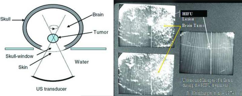
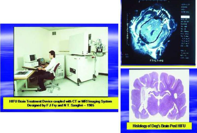
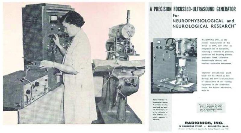
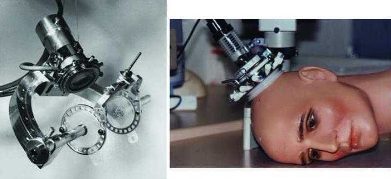
The field of magnetic resonance imaging-guided high-intensity focused ultrasound surgery (MRgFUS) is a rapidly evolving one, with many potential applications in neurosurgery. The first of 3 articles on MRgFUS, this article focuses on the historical development of the technology and its potential applications in modern neurosurgery. The evolution of MRgFUS has occurred in parallel with modern neurological surgery, and the 2 seemingly distinct disciplines share many of the same pioneering figures. Early studies on focused ultrasound treatment in the 1940s and 1950s demonstrated the ability to perform precise lesioning in the human brain, with a favorable risk-benefit profile. However, the need for a craniotomy, as well as the lack of sophisticated imaging technology, resulted in limited growth of high-intensity focused ultrasound for neurosurgery. More recently, technological advances have permitted the combination of high-intensity focused ultrasound along with magnetic resonance imaging guidance to provide an opportunity to effectively treat a variety of central nervous system disorders. Although challenges remain, high-intensity focused ultrasound-mediated neurosurgery may offer the ability to target and treat central nervous system conditions that were previously extremely difficult to address. The remaining 2 articles in this series will focus on the physical principles of modern MRgFUS as well as current and future avenues for investigation.
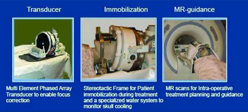

Figures

Figure 1
Figure 1
The Curie brothers noted that when pressure was applied to quartz crystals, and electrical current resulted. Converesely, if the current was an alternating voltage, the crysal oscillated with a frequency that correlated with that of the drive current. This piezoelectric phenomenon was the foundation for studies on ultrasonic energy.
Figure 2
Figure 2
Schematic demonstrating the principle of focused ultrasound-mediated tissue distruction. (Left Panel) The source is a planar ultrasound transducer situated outside of the tissue of interest. When the ultrasound beam is focused at the desired depth inside the tissue by a focusing body by a focusing lens, (Right Panel) a highly conformal dose of energy is delivered. The ultrasound beam itself can be shaped either by manipulating the lens, or by mechanical or electrical alterations to the transducers.
Figure 3
Figure 3
(Left Panel) Lynn and Putnam's ultrasound generator, placed on a cat's head. (Right Panel) Cerebellar tissue taken after 45-seconds of focused ultrasound treatement reveals a well-demarcated, wedge-shaped region of tissue damage. (Reproduced with permission)(37)
Figure 4
Figure 4
(Left panel) William Fry, the founder of the Bioacoustics lab at the University of Illinois, Champaign, with the early 4-beam HIFU applicator for neurosurgery circa 1960. (Right panel) Frank Fry receiving the Distinguished Pioneer Award from the International Society of Therapeutic Ultrasound in 2002.
Figure 5
Figure 5
An early b-mode image guided HIFU system designed in the 1970's by the Fry brothers to treat brain cancer patients.
Figure 6
Figure 6
Schematic (Left panel) HIFU-mediated glioma treatment after craniectomy, as described by Heimberger and Fry. (Right panel) Using real-time imaging with b-mode ultrasound guidance (a device known as the ‘Candy Machine’), Heimberger and Fry were able to identify a glioma, and precisely lesion desired target. (Reproduced with Permission from Focus surgery Inc)(22)
Figure 7
Figure 7
(Left panel) The more advanced Fry HIFU apparatus introduced in the 1980's, which was coupled to CT- and later MR-guidance. (Upper Right panel) MR-imaging of the dog brain following HIFU-lesioning in 1989 by Fry et al. revealed 3 single lesions on the left side of the brain, and one integrated lesion on the right. (Lower Right panel) Histology confirmed discrete, targeted lesion formation. (Photograph Courtesy of Narendra T. Sanghvi)
Figure 8
Figure 8
(Left Panel) The Cosman HIFU neurosurgical machine. Note that the transducer was placed on a modified milling machine, which allowed for rigid support and accurate positioning. (Reproduced with permission)(6) (Right Panel) Radionics advertisement from the 1950's depicting the Cosman HIFU machine. (Reproduced with permission from the JNS publishing group)
Figure 9
Figure 9
(Left panel) the Leksell frame for HIFU treatment. (Right panel) The Leksell ultrasound transducer. (Photograph courtesy of Dan Leksell. Elekta Corporation)
Figure 10
Figure 10
Components of the Insightec 4000, MR-guided focused ultrasound device. (Courtesy of Insightec Inc.)

References

    1. Ballantine HT, Jr., Bell E, Manlapaz J. Progress and problems in the neurological applications of focused ultrasound. J Neurosurg. 1960;17:858–876. - PubMed
    1. Briquard P. Paul Langevin. Ultrasonics. 1972;10:213–214. - PubMed
    1. Cartwright JH, Gonzalez DL, Piro O. Pitch perception: a dynamical-systems perspective. Proc Natl Acad Sci U S A. 2001;98:4855–4859. - PMC - PubMed
    1. Cippitelli M, Fionda C, Di Bona D, Di Rosa F, Lupo A, Piccoli M, Frati L, Santoni A. Negative regulation of CD95 ligand gene expression by vitamin D3 in T lymphocytes. J Immunol. 2002;168:1154–1166. - PubMed
    1. Cohen ZR, Zaubermann J, Harnof S, Mardor Y, Nass D, Zadicario E, Hananel A, Castel D, Faibel M, Ram Z. Magnetic resonance imaging-guided focused ultrasound for thermal ablation in the brain: a feasibility study in a swine model. Neurosurgery. 2007;60:593–600. discussion 600. - PubMed

MeSH terms