Porcine sclera as a model of human sclera for in vitro transport experiments: histology, SEM, and comparative permeability
- PMID: 19190734
- PMCID: PMC2633461
Porcine sclera as a model of human sclera for in vitro transport experiments: histology, SEM, and comparative permeability
Abstract
Purpose: To evaluate porcine sclera as a model of human sclera for in vitro studies of transscleral drug delivery of both low and high molecular weight compounds.
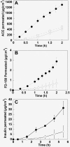
Methods: Human and porcine scleras were characterized for thickness and water content. The tissue surface was examined by scanning electron microscopy (SEM), and the histology was studied with hematoxylin-eosin staining. Comparative permeation experiments were performed using three model molecules, acetaminophen as the model compound for small molecules; a linear dextran with a molecular weight of 120 kDa as the model compound for high molecular weight drugs; and insulin, which was chosen as the model protein. Permeation parameters such as flux, lag time, and permeability coefficient were determined and compared.
Results: Human and porcine scleras have a similar histology and collagen bundle organization. The water content is approx 70% for both tissues while a statistically significant difference was found for the thickness, porcine sclera being approximately twofold thicker than human sclera. Differences in thickness produced differences in the permeability coefficient. In fact, human sclera was found to be two to threefold more permeable toward the three molecules studied than porcine sclera.
Conclusions: The results obtained in the present paper prove that porcine sclera can be considered a good model for human sclera for in vitro permeation experiments of both low and high molecular weight compounds. In fact, if the different tissue thickness is taken into account, comparable permeability was demonstrated. This suggests a possible use of this model in the evaluation of the transscleral permeation of new biotech compounds, which currently represent the most innovative and efficient therapeutic options for the treatment of ocular diseases.
Figures


References
-
- Ambati J, Adamis AP. Transscleral drug delivery to the retina and choroid. Prog Retin Eye Res. 2002;21:145–51. - PubMed
-
- Maurice DM, Polgar J. Diffusion across the sclera. Exp Eye Res. 1977;25:577–82. - PubMed
-
- Olsen TW, Sanderson S, Feng X, Hubbard WC. Porcine sclera: thickness and surface area. Invest Ophthalmol Vis Sci. 2002;43:2529–32. - PubMed
-
- Amaral J, Fariss RN, Campos MM, Robison WG, Jr, Kim H, Lutz R, Becerra SP. Transscleral-RPE permeability of PEDF and ovalbumin proteins: implications for subconjunctival protein delivery. Invest Ophthalmol Vis Sci. 2005;46:4383–92. - PubMed
MeSH terms
Substances
LinkOut - more resources
Full Text Sources
Medical
